
Page 36 - Read Online
P. 36
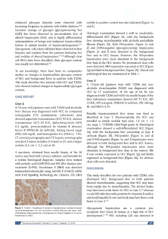
enhanced  glycogen  deposits  were  observed  with   activity in positive control was also indicated [Figure 1a
                                                         [5,6]
            increasing frequency in patients with brittle diabetes.     and b].
            Excessive storage of glycogen [glycogen-storing foci
            (GSF)] has been observed in pre-neoplastic foci of   Histologic examination showed a well- to moderately-
            altered hepatocytes (FAH), and in highly differentiated   differentiated  HCC  [Figure  2a],  with the  background
            subpopulations of benign and malignant hepatocellular   liver showing steatohepatitis with alcoholic pericellular
            lesions in animal models of hepatocarcinogenesis.    fibrosis [Figure 2b]. Both PAS-positive [Figure 2c and
                                                         [7-9]
            Glycogenotic cells (clear cell) have been observed in liver   d] and D-PAS-negative (glycogen-storing) hepatocytes
            biopsies and explants from the patients harboring foci   [Figure 2e and f] were detected in the background
            and nodules of altered hepatocytes. [10,11]  Although clear   liver and in HCC tissues. However, the PAS-positive
            cell HCCs have been described, their glycogen content   hepatocytes were more abundant in the background
            was usually not determined. [12]                   liver than in the HCC tissues. No pronounced clear cells
                                                               were detected. HK-II expression was weak in HCC [Figure
            To our knowledge, there have been no comparative   2g] and faint in background liver [Figure 2h]. Clinical and
            studies on changes in hepatocellular glycogen content   pathological data are summarized in Table 1.
            of HCC and background livers in patients with T2DM.
            This study describes two patients with HCC and T2DM,   Case 2
            who showed marked changes in hepatocellular glycogen   A  73-year-old  Japanese  man  with  T2DM  and  non-
            content.                                           alcoholic  steatohepatitis  (NASH)  was  diagnosed  with
                                                               HCC  by  CT  examination.  At  the  age  of  64,  he  was
            CASE REPORT                                        diagnosed with T2DM and NASH via needle biopsy of the
                                                               liver. Laboratory examination showed AST 51 IU/L, ALT
            Case 1                                             22 IU/L, AFP 4.4 ng/mL, PIVKA-II 22 mAU/mL, FBS 140 mg/
            A 72-year-old Japanese man with T2DM and alcoholic   dL, and HbA1c 6.3%.
            liver disease was diagnosed with HCC by computed
            tomography  (CT) examination.  Laboratory  data    Partially, hepatectomized liver tissue was fixed as
            showed aspartate transaminase (AST) 95 IU/L, alanine   described  in  Case  1.  Macroscopically,  the  HCC  was
            transaminase  (ALT) 65 IU/L,  alpha-fetoprotein  (AFP)   revealed  as  simple  nodular  type  (size,  1.8  cm  ×  1.5
                                                               cm; stage 1, T1N0M0; Child-Pugh grade A). Histological
            8.2  ng/mL, protein-induced by vitamin K absence   examination showed a well-differentiated HCC [Figure
            factor II (PIVKA-II) 26 mAU/mL, fasting blood sugar   3a],  with  the  background  liver  presenting  as  type  B
            (FBS) 228 mg/dL, and hemoglobin A1c (HbA1c)  7.9%.   cirrhosis  [Figure  3b].  PAS-positive  [Figure  3c  and  d]
            CT arterial portography and CT hepatic arteriography   and D-PAS-negative [Figure 3e and f] hepatocytes were
            revealed 2 minor nodules (3-4 mm) at S5, and a larger   detected in both background liver and in HCC tissues,
            nodule (2.5 cm × 2.3 cm) at S8.                    although the PAS-positive hepatocytes were more
                                                               abundant in background liver than in the tumors. HK-
            A specimen, obtained from needle biopsy of the S8   II was weakly expressed in HCC [Figure 3g] and faintly
            tumor, was fixed with Carnoy’s solution, and formalin for   expressed in background liver [Figure 3h]. No obvious
            a routine histological diagnosis. Samples were stained   clear cells were detected.
            with periodic acid-Schiff (PAS) and PAS after diastase pre-
            treatment (D-PAS). Hexokinase II (HK-II) was detected   DISCUSSION
            immunohistochemically using anti-HK II (C64G5) rabbit
            mAb (Cell Signaling Technology, Inc. Danvers, US). HK-II   This study describes the two patients with T2DM, who
                                                               developed HCC. Background liver in both patients
                                                               showed steatohepatitis, suggesting that HCC may have
                                                               been mainly due to steatohepatitis. The alcohol intake
                                                                                                     [2]
                                                               may have been a risk factor for HCC in Case 1,  whereas
                                                               occult HBV infection with positivity for hepatitis B surface
                                                               anti-body/hepatitis B core anti-body may have been a risk
                                                               factor in Case 2. [13]

            Figure 1: Control - hexokinase II activity in hepatocellular carcinoma tissues   Glycogenotic hepatocytes are a common pre-
            (a) and background liver (b) of positive control (65-year-old male, well-  neoplastic liver lesion in human at a high risk of HCC
            differentiated adenocarcinoma in background of chronic hepatitis C) (a and b:
            hexokinase II, ×400)                               development. [11,14]  FAH, including GSF, was detected in


                 Hepatoma Research | Volume 2 | Issue 1 | January 15, 2016                                 27