
Page 291 - Read Online
P. 291
Butt et al. TAE for ruptured HCC in Pakistan
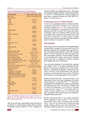
Table 1: Demographic and clinic-pathological hospital mortality was significantly lower in TAE group
characteristics of all HCC patients at baseline (n = 24) as compared to patients treated conservatively (30.8%
Characteristics Data, mean ± SD or n (%) vs. 72.7%, P = 0.04). Moreover, 30-day mortality was
Age (years) 58.29 ± 15.26 (range 17-93) also lower in patients treated with TAE (38.5% vs.
Etiology of CLD 90.9%, P = 0.01) [Table 3].
HCV 19 (79.2)
HBV 3 (12.5) Predicting factors for 30-day mortality
NBNC 2 (8.3)
Child class To find out the predicting factors for 30-day mortality,
A 0 (0) biologically plausible variables were tested on
B 16 (66.7) univariate analysis [Table 4]. The only factors which
C 8 (33.3) were found significant on univariate and multivariate
Abdominal pain
Yes 20 (83.3) analysis were TAE to control HCC bleed and control
No 4 (16.7) of bleeding. Those who underwent TAE had lower risk
Abdominal distension of mortality than conservatively treated group (OR
Yes 16 (66.7) 0.25, 95% CI 0.07-0.90, P = 0.03). Failure to control
No 8 (33.3)
Anemia bleeding was associated with higher 30-day mortality
Yes 20 (83.3) (OR 2.14, 95% CI 1.24-3.68, P = 0.009).
No 4 (16.7)
Hypovolemic shock DISCUSSION
Yes 6 (25)
No 18 (75)
Hemoperitonium In this study, we have evaluated the clinicopathological
Yes 13 (54.2) characteristics, treatment outcomes and survival of
No 11 (45.8) patients presenting with spontaneously ruptured HCC
Mean hemoglobin (g/dL) 8.4 ± 3.0 who were treated conservatively or with TAE. Success
9
Platelet count (10 /L) 202.58 ± 176.50
9
Total lecucocyte count (10 /L) 10.96 ± 4.17 rate for control of bleeding via TAE was higher than
Prothrombin time (s) 17.38 ± 5.64 with conservative treatment. Overall median duration
Mean creatinine (mg/dL) 1.35 ± 0.57 of survival after HCC rupture was longer for patients
Serum total bilirubin (mg/dL) 3.52 ± 2.87 treated with TAE. In-hospital and 30-day mortality
Alanine transaminase (IU/L) (median) 50.00 (range 13-768) were significantly lower in TAE group.
Alkaline phosphatase (IU/L) 210.13 ± 158.07
Albumin (g/dL) 2.36 ± 0.54 The reported prevalence of spontaneously ruptured
Tumor size (size of largest lesion in cm) 7.76 ± 4.22 (1.7-17.7) HCC ranges 5-15%. [18] The exact mechanism and risk
AFP (IU/mL) (median) 52.00 (range 1.00-100000) factors for spontaneous rupture are not well known.
Macroscopic types
Solitary 3 (12.5) However, subcapsular localization, rapid growth with
Paucifocal (≤ 3 nodules) 3 (12.5) tumor necrosis, portal hypertension and regional
Multifocal (> 3 nodules) 12 (50.0)
Massive (huge diameter > 10 cm, 6 (25.0) increase of venous pressure due to tumor thrombi or
undefined boundaries)/infiltrative direct invasion could be responsible for HCC rupture. [19]
Hepatic lobes (location of rupture)
Right 12 (50.0) Sudden abdominal pain, hemoperitoneum and
Left 1 (4.2)
Both 11 (45.8) hypovolemic shock have been reported as the typical
Stage of HCC clinical features of ruptured HCC. [6,9,20] Moreover,
Non-advanced 3 (12.5) hemoperitoneum ascertained by performing abdominal
Advanced 21 (87.5) paracentesis has been considered a reliable test
PVT
Yes 10 (41.7) to confirm the diagnosis in up to 86% of clinically
No 14 (58.3) suspected HCC rupture. [21] Consistent with the
Extra hepatic spread results of other studies most of our patients were
Yes 9 (37.5) male, presented with abdominal pain and distention,
No 15 (62.5)
hemoperitoneum and shock.
HCC: hepatocellular carcinoma; CLD: chronic liver disease; HCV:
hepatitis C virus; HBV: hepatitis B virus; NBNC: non-B, non-C;
MELD: Model for End Stage Liver Disease; AFP: alfa fetoprotein; Doppler ultrasound and CT are useful modalities
PVT: portal vein thrombosis for the diagnosis of HCC rupture. [22] The CT scan
demonstrate HCC rupture by showing the vascular
TAE group (39 days, interquartile range 88 days) as tumor, extent of the bleed and by showing serial
compared to conservatively treated group (5 days, density changes with the age of the hematoma. [23,24]
interquartile range 10 days) (P = 0.03). In addition, in- Triphasic contrast enhanced CT scan was done for all
282 Hepatoma Research ¦ Volume 2 ¦ October 21, 2016