
Page 264 - Read Online
P. 264
Morise et al. Repeat LLR for recurrent HCC in cirrhotic liver
A A B
B
C D
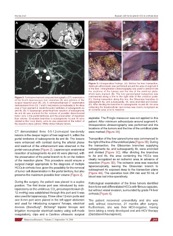
Figure 3: Intraoperative findings. (A): Before the liver transection,
minimum adhesiolysis was performed around the area of segment 4
of the liver. Intraoperative ultrasonography was used to demonstrate
the locations of the tumors and the line of the umbilical plate,
which were marked. (B): The liver parenchymal transection was
Figure 2: Contrast-enhanced computed tomography (CT) examination commenced along a line to the right side of the umbilical plate.
at the fourth laparoscopic liver resection (A) and schema of the (C): During transection along this line, the Glissonian branches to
surgical resection plan (B). (A): A contrast-enhanced CT examination subsegment 4a, and subsequently, 4b, were encircled and divided.
demonstrated three (12, 7 and 5 mm) lesions (arrowheads) in the deep (D): After dividing the branches to subsegments 4a and 4b, the area
area of liver segment 4, inside the portal territories of subsegments 4a containing the hepatocellular carcinomas was clearly recognized as
and 4b. (B): A laparoscopic anatomical liver resection of subsegments an ischemic area, prior to resection
4a and 4b was planned for the removal of possible disseminated
tumor cells in the portal territories and the preservation of maximum
liver volume. Glissonian branches to subsegments 4a and 4b were aspirator. The Pringle maneuver was not applied to this
divided at their roots (bars), while 4c was preserved on the bottom of patient. After minimum adhesiolysis around segment 4,
the resection plane (arrow). White circles indicate tumors intraoperative ultrasonography was performed and the
locations of the tumors and the line of the umbilical plate
CT demonstrated three 0.5-1.2-cm-sized low-density were marked [Figure 3A].
lesions in the deeper region of liver segment 4, within the
portal territories of subsegments 4a and 4b. The lesions Transection of the liver parenchyma was commenced to
were enhanced with contrast during the arterial phase the right of the line of the umbilical plate [Figure 3B]. During
and washout of the enhancement was observed in the the transection, the Glissonian branches supplying
portal-venous phase [Figure 2]. Laparoscopic anatomical subsegments 4a, and subsequently 4b, were encircled
resection of subsegments 4a and 4b were planned, with and divided [Figure 3C]. After dividing the branches
the preservation of the portal branch to 4c on the bottom to 4a and 4b, the area containing the HCCs was
of the resection plane. This procedure would ensure a clearly recognized as an ischemic area, in advance of
surgical margin appropriate to the diagnosis of multiple resection [Figure 3D]. The ischemic area was resected
HCCs in cirrhotic liver, given the possibility for the removal laparoscopically, leaving the Glissonian branch to
of tumor cell dissemination in the portal territory, but also subsegment 4c exposed deep to the transection plane
[Figure 4A]. The operation took 284 min and 50 mL of
preserve the maximum possible liver volume [Figure 2]. blood was lost intra-operatively.
During the surgery, the patient was placed in a supine Pathological examination of the three tumors identified
position. The first trocar port was introduced by mini- them to be well-differentiated HCCs with fibrous capsules,
laparotomy on the umbilicus; CO -pneumoperitonium (8- but without vessel invasion, surrounded by grade F4 liver
2
12 mmHg) was established through this port and it was cirrhosis [Figure 4].
also used for laparoscopy. Three other 12-mm ports and
one 8-mm port were placed in the left upper abdomen The patient recovered uneventfully and she was
and used for introducing surgeons’ forceps, electrical well, without recurrence, 21 months after surgery.
devices (SonoSurg , BiClamp bipolar forceps and Furthermore, she was then HCV-negative, having
®
®
irrigation monopolar electrical cautery using soft-mode been taking a newly developed oral anti-HCV therapy
coagulation), clips and a Cavitron ultrasonic surgical (Daclatasvir/Asunaprevir).
Hepatoma Research ¦ Volume 2 ¦ September 19, 2016 255