
Page 263 - Read Online
P. 263
Morise et al. Repeat LLR for recurrent HCC in cirrhotic liver
INTRODUCTION of repeat LLR for the treatment of HCC. [22,23]
Since the first successful report of laparoscopic CASE REPORT
liver wedge resection in 1991, laparoscopic liver
[1]
resection (LLR) has been thought to be a “less A 73-year-old woman with hepatitis C virus (HCV)-
invasive” procedure than open liver resection. Use related liver cirrhosis (LC) was admitted to our
of this technique is especially beneficial for patients department for treatment of three lesions in liver
with concurrent hepatocellular carcinoma (HCC) and segment 4. These were revealed by contrast-enhanced
chronic liver disease (CLD). [2-4] However, accumulated computed tomography (CT) examination undertaken
experience of this technique and technological during the follow up to three LLRs that were performed
developments have facilitated the expansion of the 73, 45, 23 months previously [Figure 1]. The patient
indications for LLR. [5-7] It is becoming clear that the had no history of hepatic encephalopathy, ascites
magnified caudal view offered by laparoscopy allows (except immediately postoperatively) and no specific
improved visualization, especially for the hilar and treatment history except that of the liver disease.
dorsal area of the liver, and is thus beneficial for the
dissection of hilar Glissonian pedicles and the inferior The laboratory data showed decreased white blood cell
vena cava (IVC). [7-9] LLRs of major hepatectomy and platelet counts (1,800 and 68,000/µL, respectively)
and, even, with combined resection of major hepatic and plasma albumin (3.5 g/dL) and mild elevations in
veins are now increasingly reported, [10-12] despite the plasma aspartate transaminase (AST, 76 IU/L) and
latter previously being a contraindication. Reports of alanine transaminase (ALT, 71 IU/L). The prothrombin
repeated LLR procedures [13-16] are also increasing. time (78%), plasma levels of total bilirubin (0.6 mg/dL) and
However, these reports have generally included both prothrombin induced by vitamin K absence-II (PIVKA-
cases of HCC with CLD and of metastatic disease II, 9 mAU/mL) were within their normal ranges, but
without background liver disease. [17-21] The indication alpha-fetoprotein (AFP) showed a mild elevation (to
and efficacy of repeated LLR for HCC in a setting of CLD 67.5 ng/mL). The 15-min value during the clearance
alone has yet to be fully determined. Here we present rate of indocyanine green loading test (ICG-R15) was
a case report of a fourth LLR for recurrent HCCs in 24.1%; this had not deteriorated over the 73 months
cirrhotic liver and review the previously reported cases since the first LLR [Table 1].
Table 1: Perioperative clinical variables associated with each LLR
1st 2nd 3rd 4th
ICG-R15 20.9 27.5 27.0 24.1
Bleeding (mL) 35 30 NC 50
Operating time (min) 288 168 216 274
POHS (days) 11 9 9 8
LLR: laparoscopic liver resection; ICG-R15: 15 min value during the clearance rate of indocyanine green loading test; 1st: ICG-R15 and
perioperative course of first LLR; 2nd: ICG-R15 and perioperative course of second LLR; 3rd: ICG-R15 and perioperative course of third LLR; 4th:
ICG-R15 and perioperative course of fourth LLR; NC: low, unquantifiable; POHS: postoperative hospital stay
A B C
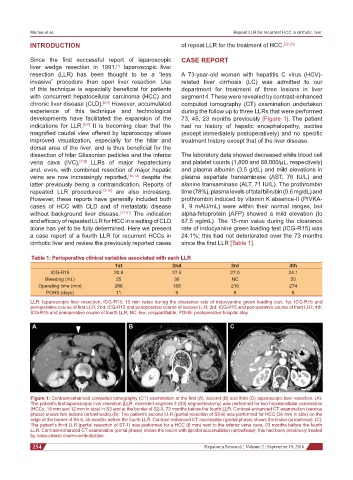
Figure 1: Contrast-enhanced computed tomography (CT) examination at the first (A), second (B) and third (C) laparoscopic liver resection. (A):
The patient’s first laparoscopic liver resection [LLR, extended segment 3 (S3) segmentectomy] was performed for two hepatocellular carcinomas
(HCCs, 18 mm and 12 mm in size) in S3 and at the border of S2-3, 73 months before the fourth LLR. Contrast-enhanced CT examination (venous
phase) shows two lesions (arrowheads).(B): The patient’s second LLR (partial resection of S5-6) was performed for HCC (30 mm in size) on the
edge of the border of S5-6, 45 months before the fourth LLR. Contrast-enhanced CT examination (portal phase) shows the lesion (arrowhead). (C):
The patient’s third LLR (partial resection of S7-1) was performed for a HCC (8 mm) next to the inferior vena cava, 23 months before the fourth
LLR. Contrast-enhanced CT examination (portal phase) shows the lesion with lipiodol accumulation (arrowhead); this had been previously treated
by trans-arterial chemo-embolization
254 Hepatoma Research ¦ Volume 2 ¦ September 19, 2016