
Page 175 - Read Online
P. 175
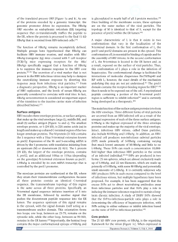
of the translated precore ORF [Figure 1a and b]. As one   is glycosylated in nearly half of all S-protein moieties. [39]
            of the  proteins  encoded by  a  genomic  transcript, the   Once budding of the membrane occurs, these epitopes
            genomic  promoter  drives  its  expression.  The  HBeAg   are  on  the  outer  surface  of  the  viral  particles.  The
            ORF  encodes an endoplasmic reticulum  (ER) targeting   topology of M is  identical to that of S,  except for the
            sequence  that co-translationally traffics the  peptide to   presence of preS2 within the ER lumen. [40]
            the ER, where the protein is processed to the final 15 kD
            HBeAg that is secreted from HBV-infected cells. [34]  A major characteristic of L is that it exists in two
                                                              conformations that vary in the localization of the
            The function of HBeAg remains incompletely defined.   N-terminal  domain.  In  the  first  conformation of L,  the
            Multiple  groups  have  hypothesized  that  HBeAg  can   preS1 and preS2 domains are present in the cytosol. This
            facilitate HBV immune  evasion,  and studies  with  HBc/  conformation of L is essential for binding of capsids and for
            HBeAg-transgenic (tg) mice crossed with T cell receptor   the assembly of HBV virions. In the second conformation
            (TCR)-Tg  mice  expressing  receptors for the  HBc/  of L, the N-terminus is located in the ER lumen and, as
            HBeAgs specifically suggest  that a function of HBeAg   a result, exposed on the surface of viral particles. Thus,
            is to suppress the immune  response to the HBV core   this  conformation of L  plays a role in  the  infection of
            protein. [35,36]  The secretion of a viral marker that is not   hepatocytes. The conformational change is facilitated by
            present in the HBV infectious virion may help to dampen   interactions of molecular chaperones Hsc70/Hsp40 and
            the neutralizing immune response by diverting this   BiP  with  L;  however, the exact details of the mechanism
            response away from infectious viral particles.  From   underlying this step are not yet understood.  The preS1
                                                    [11]
                                                                                                  [11]
            a diagnostic perspective, HBeAg is an important marker   domain contains the receptor-binding region for HBV, [41,42]
            of HBV replication, and the levels of serum HBeAg are   thus it needs to be exposed out of the cell. A myristylated
            generally considered to correlate with viral titer. In fact,   peptide containing a portion of the N-terminal preS1
                                                                                              [41]
            HBeAg seroconversion is considered an important aspect   region is sufficient to inhibit infection  and is currently
            of the transition to the inactive carrier state of infection   being developed as a therapeutic. [43]
            (described below). [37]
                                                              The main function of the surface antigen proteins is to form
            Surface antigens                                  the HBV envelope. Three different forms of viral particles
            HBV encodes three envelope proteins, or surface antigens,   are secreted from an HBV-infected cell as a result of the
            that make up the viral envelope: large (L), middle (M), and   unequal expression of each of the three surface antigens.
            small (S) surface antigen [Figure 1a and c]. The smallest   S-HBsAg is the highest expressed of the three envelope
            envelope protein, or S (24 kD), is 226 amino acids (aa) in   proteins and makes up the majority of the viral envelope.
            length and makes up a shared C-terminal region of the two   Intact,  infectious  HBV  virions,  called Dane  particles,
            longer envelope proteins. The M protein (31 kD) contains   also include M-HBsAg and L-HBsAg. In addition, an HBV-
            the S sequence with a 55aa N-terminal extension known   infected cell produces non-infectious subviral particles
            as preS2. Expression of the M- and S-encoding mRNA is   (SVP) made primarily  of S-HBsAg containing varying
            driven by the S promoter, with translation initiating from   (but much lower) amounts of M-HBsAg and little to no
            an upstream (M) or downstream (S) AUG. The L protein   L-HBsAg. These SVPs can reach a concentration 10,000-
            (39 kD), the largest of the envelope proteins, contains   fold higher  than infectious HBV particles in the serum
            S, preS2, and an additional 108aa or 119aa (depending   of an infected individual. [44,45]  SVPs are produced in two
            on the genotype) N-terminal extension known as preS1.   forms: 25 nm spheres, which are almost exclusively made
            L-HBsAg is encoded by its own mRNA transcript that is   up of S-HBsAg, and 22 nm filaments, which are made up
            controlled by the preS1 promoter.                 primarily of S-HBsAg, with some M-HBsAg and potentially
                                                              small amounts of L-HBsAg. It is currently unknown why
            The envelope proteins are synthesized at the ER, where   HBV produces SVPs in such excess compared to the level
            they attain their transmembrane configuration. Because   of infectious virions, but multiple hypotheses have been
            all three proteins contain an identical C-terminal   proposed. For example, it has been suggested that the
            sequence,  the transmembrane  topology of this  region   excess SVPs act to divert neutralizing  antibodies away
            is  the  same  across all three  proteins.  Specifically,  an   from infectious particles and that SVPs  play a role in
            N-terminal signal sequence initiates insertion of S into   inducing the immune tolerance required to sustain a long-
            the  ER  membrane,  followed by  another signal  that   term  chronic infection. A study of DHBV SVPs  showed
            pushes the downstream peptide sequence into the ER   that  the  SVP-to-infectious-particle  ratio  plays a  role in
            lumen.  The sequence  upstream  of this  signal  remains   determining the efficiency of hepatocyte infection, with
            in the cytosol, with the signal domain itself acting as a   SVPs acting to either enhance or inhibit infection based
            transmembrane anchor domain. This orientation forms   on the ratio of SVP-to-infectious-particles. [46]
            two loops; one loop, between aa 23-79, remains on the
            cytosolic side, while the other loop, between aa 99-169,   Core protein
            remains in the ER lumen. [38]  Importantly, the luminal loop   The 21 kD HBV core protein, or HBcAg, is the organizing
            contains the major conformational epitope of HBsAg and   framework for the virion [Figure 1c]. When expressed in
            166                                                           Hepatoma Research | Volume 2 | July 1, 2016