
Page 50 - Read Online
P. 50
a b
a b
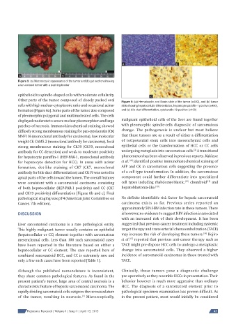
Figure 5: (a) Macroscopic appearance of the tumor and (b) gut section showing
a tan-colored tumor with a pushing border
epithelioid to spindle-shaped cells with moderate cellularity. c
Other parts of the tumor composed of closely packed oval Figure 6: (a) Hematoxylin and Eosin slide of the tumor (×400), and (b) tumor
cells with high nuclear-cytoplasmic ratio and occasional acinar slide showing hepatocellular differentiation, hepatocyte paraffi n-1 positive (×400),
formation [Figure 6a]. Some parts of the tumor also composed and (c) bile duct differentiation, cytokeratin 19 positive (×400)
of pleomorphic polygonal and multinucleated cells. The cells
displayed moderate to severe nuclear pleomorphism and large malignant epithelioid cells of the liver are found together
patches of necrosis. Immunohistochemical staining showed with pleomorphic spindle-cells diagnostic of sarcomatous
diffusely strong membranous staining for pan-cytokeratin (CK) change. The pathogenesis is unclear but most believe
MNF116 (monoclonal antibody for carcinoma), low molecular that these tumors are as a result of either a differentiation
weight CK CAM5.2 (monoclonal antibody for carcinoma), focal of totipotential stem cells into mesenchymal cells and
strong membranous staining for CK19 (CK19, monoclonal epithelial cells or the transformation of HCC or CC cells
[2]
antibody for CC detection) and weak to moderate positivity undergoing metaplasia into sarcomatous cells. A transitional
for hepatocyte paraffin-1 (HEP-PAR-1, monoclonal antibody phenomenon has been observed in previous reports. Kakizoe
[10]
for hepatocyte detection for HCC). In areas with acinar et al. identified positive immunohistochemical staining of
formation, dot-like staining of CK7 (CK7, monoclonal AFP and CK in sarcomatous cells suggesting the presence
antibody for bile duct differentiation) and CK19 was noted in of a cell type transformation. In addition, the sarcomatous
apical parts of the cells toward the lumen. The overall features component could further differentiate into specialized
[12]
[11]
were consistent with a sarcomatoid carcinoma consisting cell types including rhabdomyoblastic, chondroid and
of both hepatocellular (HEP-PAR-1 positivity) and CC (CK7 hepatoblastoma-like. [13]
and CK19 positivity) differentiation [Figure 6b and c]. Final
pathological staging was pT4 (American Joint Committee on No definite identifiable risk factor for hepatic sarcomatoid
Cancer, 7th edition). carcinoma exists so far. Previous series reported an
approximately 50% HBV infection rate in these tumors. There
DISCUSSION is however, no evidence to suggest HBV infection is associated
with an increased risk of their development. It has been
Liver sarcomatoid carcinoma is a rare pathological entity. suggested that previous cancer treatment including systemic
This highly malignant tumor usually contains an epithelial target therapy and trans-arterial chemoembolization (TACE)
[14]
(hepatocellular or CC) element together with sarcomatous may increase the risk of developing these tumors. Kojiro
mesenchymal cells. Less than 100 such sarcomatoid cases et al. reported that previous anti-cancer therapy such as
[15]
have been reported in the literature based on either a TACE might pre-dispose HCC cells to undergo a metaplastic
hepatocellular or CC element. The case reported here of change into sarcomatoid cells. They observed a higher
combined sarcomatoid HCC, and CC is extremely rare and incidence of sarcomatoid carcinomas in those treated with
only a few such cases have been reported [Table 1]. TACE.
Although the published nomenclature is inconsistent, Clinically, these tumors pose a diagnostic challenge
they share common pathological features. As found in the pre-operatively as they resemble HCCs in presentation. Their
present patient’s tumor, large area of central necrosis is a behavior however is much more aggressive than ordinary
characteristic feature of hepatic sarcomatoid carcinoma. The HCC. The diagnosis of a sarcomatoid element prior to
rapidly dividing sarcomatoid cells outgrow the neovasculature pathological specimen examination has proven difficult. As
of the tumor, resulting in necrosis. Microscopically, in the present patient, most would initially be considered
[1]
Hepatoma Research | Volume 1 | Issue 1 | April 15, 2015 43