
Page 49 - Read Online
P. 49
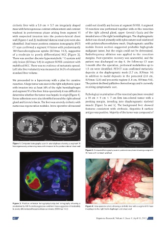
cirrhotic liver with a 5.9 cm × 5.7 cm irregularly shaped   could not identify any lesions at segment IV/VIII. A segment
          mass with heterogeneous contrast enhancement and contrast   VI resection was performed together with en bloc resection
          washout in portovenous phase arising from segment VI   of the right adrenal gland, upper Gerota’s fascia and the
          with suspected invasion into the postero-lateral chest   invaded area of the right hemidiaphragm. The diaphragmatic
          wall [Figures 1 and 2]. Incidental bilateral renal cysts were also   defect was closed primarily with nylon sutures and reinforced
          identified. Dual tracer positron emission tomography (PET)   with polytetrafluoroethylene mesh. Diaphragmatic satellite
          CT scan confirmed a segment VI lesion with predominantly   nodule frozen section suggested probable high-grade
          18F-fluorodeoxyglucose uptake (SUVmax 14.2), suggestive   malignant tumor, but the origin could not be determined.
          of a moderate to poorly differentiated HCC [Figure 3].   Radiofrequency ablation was applied to the resection
                                              11
          There was another discrete hypermetabolic  C-acetate avid   margins. Post-operative recovery was uneventful, and the
          only lesion (SUVmax 9.8) in segment IV/VIII consistent with   patient was discharged on day 6. On follow-up CT scan
          multifocal HCC. There was no evidence of metastatic spread.   1-month after the operation, peritoneal nodularities up to
          Left lobe liver volumetry was measured at 34.5% of estimated   1.5 cm were identified. PET/CT scan confirmed metastatic
          standard liver volume.                              deposits at the diaphragmatic mesh (2.7 cm, SUVmax 14)
                                                              in addition to nodal deposits in the paracaval (2.8 cm,
          We proceeded to a laparotomy with a plan for curative   SUVmax 12.6) and pre-aortic regions (1.8 cm, SUVmax 9.6).
          resection. A large tumor was seen in the right subphrenic space   The patient declined palliative chemotherapy and is currently
          with invasion into at least 50% of the right hemidiaphragm   receiving symptomatic care.
          and segment VI of the liver. Intra-operatively it was difficult to
          determine whether the tumor was hepatic in origin [Figure 4].   Pathological examination of the resected specimen revealed
          Dense adhesions were also identified around the right adrenal   a 10 cm × 9 cm × 7 cm firm tan-colored tumor with a
          gland and Gerota’s fascia. The liver was severely cirrhotic with   pushing margin, invading into diaphragmatic skeletal
          numerous regeneration nodules. Intra-operative ultrasound   muscle [Figure 5a and b]. The background liver showed
                                                              features consistent with cirrhosis. Hepatitis B surface
                                                              antigen was positive. Majority of the tumor was composed of


          Figure 1: Computed tomography scan in arterial phase showing a segment VI
          heterogeneously enhancing mass with invasion to the postero-lateral chest wall
                                                              Figure 2: Computed tomography scan in portovenous phase showing a segment
                                                              VI mass with contrast washout


          Figure 3: Positron emission tomography/computed tomography showing a
          predominantly 18F-fl uorodeoxyglucose-avid liver mass suggestive of moderately   Figure 4: Intra-operative photo showing a cirrhotic liver with a segment VI mass
          to poorly differentiated hepatocellular carcinoma (SUVmax 14.2)  invading into the right hemi-diaphragm and chest wall


          42                                                          Hepatoma Research | Volume 1 | Issue 1 | April 15, 2015