
Page 48 - Read Online
P. 48
Page 6 of 16 Kościuszko et al. Hepatoma Res 2021;7:51 https://dx.doi.org/10.20517/2394-5079.2021.17
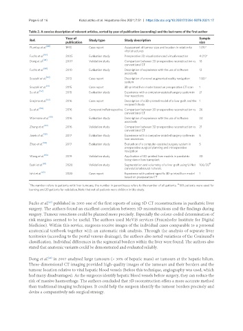
Table 2. A concise description of relevant articles, sorted by year of publication (ascending) and the last name of the first author
Year of Sample
Ref. Study type Study description
publication size
[46]
Plumley et al. 1995 Case report Assessment of tumour size and location in relation to 1 (9)*
vital structures
[47]
Fuchs et al. 2005 Evaluation study Preoperative 3D visualisation and virtual resection 4 (11)*
Dong et al. [48] 2007 Validation study Comparison between 3D preoperative reconstruction vs. 18
conventional CT
[49]
Fuchs et al. 2010 Evaluation study Description of experience with the use of software 12
assistants
[50]
Souzaki et al. 2013 Case report Description of a novel augmented reality navigation 1 (6)*
system
[51]
Souzaki et al. 2015 Case report 3D-printed liver model based on preoperative CT scan 1
Su et al. [52] 2015 Evaluation study Experience with a computer-assisted surgery system in 21
liver resections
Soejima et al. [53] 2016 Case report Description of a 3D-printed model of a liver graft and the 1
recipient’s body
Su et al. [54] 2016 ComparativeRetrospective Comparison between 3D preoperative reconstruction vs. 26
conventional CT
[55]
Warmann et al. 2016 Evaluation study Description of experience with the use of software 24
assistants
[57]
Zhang et al. 2016 Validation study Comparison between 3D preoperative reconstruction vs. 21
conventional CT
[58]
Janek et al. 2017 Evaluation study Experience with a computer-assisted surgery system in 5
liver resections
[59]
Zhao et al. 2017 Evaluation study Evaluation of a computer-assisted surgery system in 5
preoperative surgical planning and intraoperative
navigation
Wang et al. [60] 2019 Validation study Application of 3D-printed liver models in paediatric 30
living-donor liver transplant
[61] #
Esaki et al. 2020 Validation study Segmentation and volumetry of a liver graft using U-Net 100/20
convolutional neural network
[63]
Ishii et al. 2020 Case report Experience with patient-specific 3D-printed liver model 1
based on preoperative CT
* #
The number refers to patients with liver tumours; the number in parentheses refers to the number of all patients. 100 patients were used for
learning and 20 patients for validation. Note that not all patients were children in this study.
Fuchs et al. published in 2005 one of the first reports of using 3D CT reconstructions in paediatric liver
[47]
surgery. The authors found an excellent correlation between 3D reconstructions and the findings during
surgery. Tumour resections could be planned more precisely. Especially the colour-coded determination of
risk margins seemed to be useful. The authors used MeViS services (Fraunhofer Institute for Digital
Medicine). Within this service, surgeons receive images of the individual cases comparable to a personal
anatomical textbook together with an automatic risk analysis. Through the analysis of separate liver
territories (according to the portal venous drainage), the authors also noted variations of the Couinaud’s
classification. Individual differences in the segmental borders within the liver were found. The authors also
stated that anatomic variants could be demonstrated and evaluated reliably.
Dong et al. in 2007 analysed large tumours (> 30% of hepatic mass) or tumours at the hepatic hilum.
[48]
Three-dimensional CT imaging provided high-quality images of the tumours and their borders and the
tumour location relative to vital hepatic blood vessels (before this technique, angiography was used, which
had many disadvantages). As the surgeons identify hepatic blood vessels before surgery, they can reduce the
risk of massive haemorrhage. The authors concluded that 3D reconstruction offers a more accurate method
than traditional imaging techniques. It could help the surgeon identify the tumour borders precisely and
devise a comparatively safe surgical strategy.