
Page 46 - Read Online
P. 46
Page 4 of 16 Kościuszko et al. Hepatoma Res 2021;7:51 https://dx.doi.org/10.20517/2394-5079.2021.17
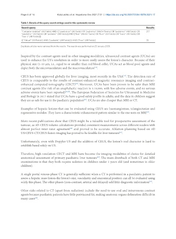
Table 1. Details of the query search strings used in this systematic review
Search query Results
“Computer-assisted” (All Fields) AND {[“paediatrics” (All Fields) OR “pediatrics” (MeSH Terms) OR “pediatrics” (All Fields) OR 217
“paediatric” (All Fields) OR “pediatric” (All Fields)] AND [“liver” (MeSH Terms) OR “liver” (All Fields) OR “livers” (All Fields) OR
“liver s” (All Fields)]}
{[“Virtual” (All Fields)] AND [“pediatric” (All Fields)]} AND [“liver” (All Fields)] 35
Duplicate articles were removed from the results. The search was performed on 25 January 2021.
Inspired by the contrast agents used in other imaging modalities, ultrasound contrast agents (UCAs) are
used to enhance the US’s resolution in order to more easily assess the lesion’s character. Because of their
physical size (1-10 μm, i.e., equal to or smaller than red blood cells), UCAs act as blood pool agents and
depict both the microvasculature and the macrovasculature .
[18]
[19]
CEUS has been approved globally for liver imaging, most recently in the USA . The detection rate of
CEUS is comparable to the results of contrast-enhanced magnetic resonance imaging and contrast-
enhanced computed tomography (CECT) . Moreover, UCAs have been proven to be safer than MRI
[20]
contrast agents (the risk of an anaphylactic reaction is 1:10,000, with few adverse events, and no serious
adverse events have been reported) [21,22] . The European Federation of Societies for Ultrasound in Medicine
and Biology in 2017 stated that UCAs have a good safety profile in adults, and the data in children suggest
they are as safe for use in the paediatric population . UCAs are also cheaper than MRI or CT.
[23]
Examples of hepatic lesions that can be evaluated using CEUS are haemangiomas, telangiectasias and
regenerative nodules. They have a characteristic enhancement pattern similar to the one seen on MRI .
[24]
More recent publications show that CEUS might be a valuable tool for preoperative assessment of the
tumour, as 3D-CEUS volume calculations provided consistent measurements across different readers with
[25]
almost perfect inter-rater agreement and proved to be accurate. Ablation planning based on 3D
US/CEUS-CT/CEUS fusion imaging has proved to be feasible for liver tumours .
[26]
Unfortunately, even with Doppler US and the addition of CEUS, the lesion’s real character is hard to
establish based solely on US.
Therefore, high-resolution CECT and MRI have become the imaging modalities of choice for detailed
[27]
anatomical assessment of primary paediatric liver tumours . The main drawback of both CT and MRI
examinations is that they both require sedation in children under 5 years old (and sometimes in older
children).
A single portal venous phase CT is generally sufficient when a CT is performed in a paediatric patient to
assess a hepatic mass lesion-the lesion’s size, vascularity and anatomical position can all be evaluated using
[28]
solely this phase. The other phases (non-contrast, arterial and delayed) add little diagnostic information .
Other risks related to CT (apart from radiation) include the need to use oral and intravenous contrast
agents because paediatric patients have little perivisceral fat, making anatomic organs delineation difficult in
many cases .
[29]