
Page 21 - Read Online
P. 21
Zhang et al. Hepatoma Res 2020;6:30 I http://dx.doi.org/10.20517/2394-5079.2020.17 Page 9 of 16
A B C
D E F
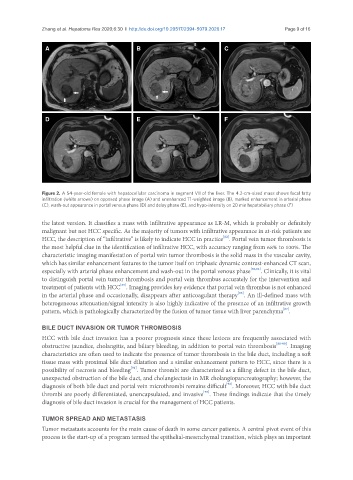
Figure 2. A 54-year-old female with hepatocellular carcinoma in segment VII of the liver. The 4.2-cm-sized mass shows focal fatty
infiltration (white arrows) on opposed phase image (A) and unenhanced T1-weighted image (B), marked enhancement in arterial phase
(C), wash-out appearance in portal venous phase (D) and delay phase (E), and hypo-intensity on 20 min hepatobiliary phase (F)
the latest version. It classifies a mass with infiltrative appearance as LR-M, which is probably or definitely
malignant but not HCC specific. As the majority of tumors with infiltrative appearance in at-risk patients are
[82]
HCC, the description of “infiltrative” is likely to indicate HCC in practice . Portal vein tumor thrombosis is
the most helpful clue in the identification of infiltrative HCC, with accuracy ranging from 68% to 100%. The
characteristic imaging manifestation of portal vein tumor thrombosis is the solid mass in the vascular cavity,
which has similar enhancement features to the tumor itself on triphasic dynamic contrast-enhanced CT scan,
especially with arterial phase enhancement and wash-out in the portal venous phase [83,84] . Clinically, it is vital
to distinguish portal vein tumor thrombosis and portal vein thrombus accurately for the intervention and
[85]
treatment of patients with HCC . Imaging provides key evidence that portal vein thrombus is not enhanced
[86]
in the arterial phase and occasionally, disappears after anticoagulant therapy . An ill-defined mass with
heterogeneous attenuation/signal intensity is also highly indicative of the presence of an infiltrative growth
[87]
pattern, which is pathologically characterized by the fusion of tumor tissue with liver parenchyma .
BILE DUCT INVASION OR TUMOR THROMBOSIS
HCC with bile duct invasion has a poorer prognosis since these lesions are frequently associated with
obstructive jaundice, cholangitis, and biliary bleeding, in addition to portal vein thrombosis [88-90] . Imaging
characteristics are often used to indicate the presence of tumor thrombosis in the bile duct, including a soft
tissue mass with proximal bile duct dilatation and a similar enhancement pattern to HCC, since there is a
[91]
possibility of necrosis and bleeding . Tumor thrombi are characterized as a filling defect in the bile duct,
unexpected obstruction of the bile duct, and cholangiectasis in MR cholangiopancreatography; however, the
[92]
diagnosis of both bile duct and portal vein microthrombi remains difficult . Moreover, HCC with bile duct
[93]
thrombi are poorly differentiated, unencapsulated, and invasive . These findings indicate that the timely
diagnosis of bile duct invasion is crucial for the management of HCC patients.
TUMOR SPREAD AND METASTASIS
Tumor metastasis accounts for the main cause of death in some cancer patients. A central pivot event of this
process is the start-up of a program termed the epithelial-mesenchymal transition, which plays an important