
Page 18 - Read Online
P. 18
Page 6 of 16 Zhang et al. Hepatoma Res 2020;6:30 I http://dx.doi.org/10.20517/2394-5079.2020.17
A B C
D E F
G
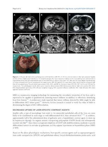
Figure 1. A 48-year-old male with a pathologically confirmed HCC with MVI. A 7.8 cm × 6.0 cm tumor in the right posterior hepatic
section shows slight hyperintensity on T2-weighted image (A), hypo-intensity relative to the surrounding liver parenchyma in
hepatobiliary phase (B), and restricted diffusion on the DWI with a b-value of 700 s/mm (C). MK map (D) shows higher signal intensity
2
of tumor compared with that of background liver parenchyma. ADC (E) and MD (F) maps show lower signal intensity compared with
2
-3
that of liver parenchyma. The calculated mean values of MK, ADC, and MK for the tumor were 0.99, 1.10 × 10 mm /s, and 1.28 mm /s,
2
respectively. The pathological results after tumor resection demonstrated HCC [Edmondson-Steiner (III) with positive MVI (G)].
HCC: hepatocellular carcinoma; DWI: diffusion-weighted imaging; ADC: apparent diffusion coefficient; MD: mean diffusion; MK: mean
apparent kurtosis coefficient
MRE is a noninvasive imaging technology for measuring the viscoelastic properties of the liver, and is
reported to be capable of quantitatively assessing tumor stiffness, in addition to effectively identifying
[39]
focal liver lesions . A preliminary study reported that tumor stiffness detected by MRE might be able
[40]
to differentiate HCC tumor grade . However, further research is needed to verify the value of MRE in
determining the degree of HCC differentiation.
DECREASED UPTAKE OF LIVER-SPECIFIC CONTRAST AGENTS
Kupffer cells, a type of macrophage that reside in the sinusoidal endothelial cells of the liver, are more
likely to be distributed in early-stage or well-differentiated HCC than advanced HCC [41,42] . In addition,
approximately half of the administered dose of gadoxetic acid, a hepatobiliary contrast agent, is taken up
by hepatocytes through the organic anionic transporting polypeptide (OATP) family and subsequently
[43]
excreted into bile . Since there is progressive depletion of OATP with dedifferentiation, moderate to poorly
differentiated tumors do not take up gadoxetate, and thus appear hypointense on hepatobiliary phase (HBP)
of MRI.
Based on the above physiological mechanisms, liver-specific contrast agents such as superparamagnetic
iron-oxide nanoparticles (SPION) and gadolinium-ethoxy benzyl-diethylenetriamine penta-acetic acid