
Page 34 - Read Online
P. 34
Afyouni et al. Hepatoma Res 2023;9:28 https://dx.doi.org/10.20517/2394-5079.2023.29 Page 3 of 14
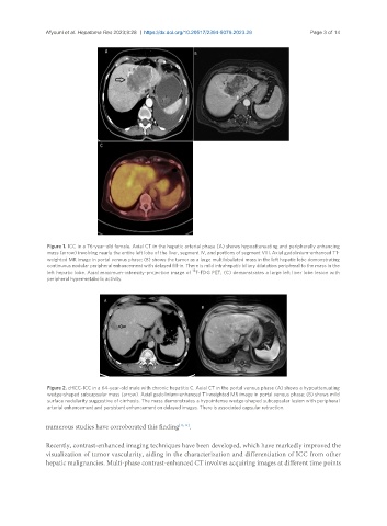
Figure 1. ICC in a 76-year-old female. Axial CT in the hepatic arterial phase (A) shows hypoattenuating and peripherally enhancing
mass (arrow) involving nearly the entire left lobe of the liver, segment IV, and portions of segment VIII. Axial gadolinium-enhanced T1-
weighted MR image in portal venous phase; (B) shows the tumor as a large multilobulated mass in the left hepatic lobe demonstrating
continuous nodular peripheral enhancement with delayed fill-in. There is mild intrahepatic biliary dilatation peripheral to the mass in the
18
left hepatic lobe. Axial maximum-intensity-projection image of F-FDG PET; (C) demonstrates a large left liver lobe lesion with
peripheral hypermetabolic activity.
Figure 2. cHCC-ICC in a 64-year-old male with chronic hepatitis C. Axial CT in the portal venous phase (A) shows a hypoattenuating
wedge-shaped subcapsular mass (arrow). Axial gadolinium-enhanced T1-weighted MR image in portal venous phase; (B) shows mild
surface nodularity suggestive of cirrhosis. The mass demonstrates a hypointense wedge-shaped subcapsular lesion with peripheral
arterial enhancement and persistent enhancement on delayed images. There is associated capsular retraction.
numerous studies have corroborated this finding [13,14] .
Recently, contrast-enhanced imaging techniques have been developed, which have markedly improved the
visualization of tumor vascularity, aiding in the characterization and differentiation of ICC from other
hepatic malignancies. Multi-phase contrast-enhanced CT involves acquiring images at different time points