


Page 29 - Read Online
P. 29
Li et al. Hepatoma Res. 2025;11:25  https://dx.doi.org/10.20517/2394-5079.2025.63  Page 7 of 17

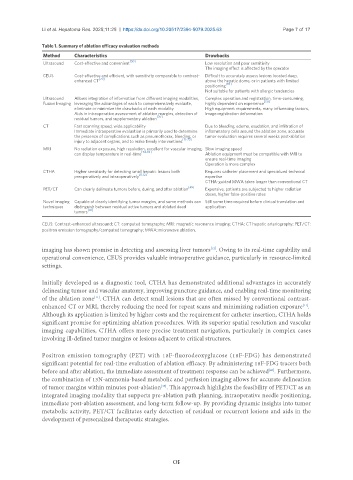
               Table 1. Summary of ablation efficacy evaluation methods
                Method    Characteristics                             Drawbacks
                Ultrasound  Cost-effective and convenient [50]        Low resolution and poor sensitivity
                                                                      The imaging effect is affected by the operator
                CEUS      Cost-effective and efficient, with sensitivity comparable to contrast-  Difficult to accurately assess lesions located deep,
                                  [70]
                          enhanced CT                                 above the hepatic dome, or in patients with limited
                                                                             [70]
                                                                      positioning
                                                                      Not suitable for patients with allergic tendencies
                Ultrasound   Allows integration of information from different imaging modalities,   Complex operation and registration, time-consuming,
                Fusion Imaging leveraging the advantages of each to comprehensively evaluate,   highly dependent on experience [50]
                          eliminate or minimize the drawbacks of each modality   High equipment requirements, many influencing factors,
                          Aids in intraoperative assessment of ablation margins, detection of   image registration deformation
                                                      [70]
                          residual tumors, and supplementary ablation
                CT        Fast scanning speed, wide applicability     Due to bleeding, edema, exudation, and infiltration of
                          Immediate intraoperative evaluation is primarily used to determine   inflammatory cells around the ablation zone, accurate
                          the presence of complications such as pneumothorax, bleeding, or   tumor evaluation requires several weeks post-ablation
                                                               [71-73]
                          injury to adjacent organs, and to make timely interventions
                MRI       No radiation exposure, high resolution, excellent for vascular imaging,  Slow imaging speed
                          can display temperature in real-time [74-76]  Ablation equipment must be compatible with MRI to
                                                                      ensure real-time imaging
                                                                      Operation is more complex
                CTHA      Higher sensitivity for detecting small hepatic lesions both   Requires catheter placement and specialized technical
                          preoperatively and intraoperatively [11,12]  expertise
                                                                      CTHA-guided MWA takes longer than conventional CT
                PET/CT    Can clearly delineate tumors before, during, and after ablation [49]  Expensive, patients are subjected to higher radiation
                                                                      doses, higher false-positive rates
                Novel imaging  Capable of clearly identifying tumor margins, and some methods can  Still some time required before clinical translation and
                techniques  distinguish between residual active tumors and ablated dead   application
                               [47]
                          tumors
               CEUS: Contrast-enhanced ultrasound; CT: computed tomography; MRI: magnetic resonance imaging; CTHA: CT hepatic arteriography; PET/CT:
               positron emission tomography/computed tomography; MWA: microwave ablation.


               imaging has shown promise in detecting and assessing liver tumors . Owing to its real-time capability and
                                                                        [12]
               operational convenience, CEUS provides valuable intraoperative guidance, particularly in resource-limited
               settings.


               Initially developed as a diagnostic tool, CTHA has demonstrated additional advantages in accurately
               delineating tumor and vascular anatomy, improving puncture guidance, and enabling real-time monitoring
                                 [11]
               of the ablation zone . CTHA can detect small lesions that are often missed by conventional contrast-
               enhanced CT or MRI, thereby reducing the need for repeat scans and minimizing radiation exposure .
                                                                                                       [11]
               Although its application is limited by higher costs and the requirement for catheter insertion, CTHA holds
               significant promise for optimizing ablation procedures. With its superior spatial resolution and vascular
               imaging capabilities, CTHA offers more precise treatment navigation, particularly in complex cases
               involving ill-defined tumor margins or lesions adjacent to critical structures.


               Positron emission tomography (PET) with 18F-fluorodeoxyglucose (18F-FDG) has demonstrated
               significant potential for real-time evaluation of ablation efficacy. By administering 18F-FDG tracers both
                                                                                            [45]
               before and after ablation, the immediate assessment of treatment response can be achieved . Furthermore,
               the combination of 13N-ammonia-based metabolic and perfusion imaging allows for accurate delineation
               of tumor margins within minutes post-ablation . This approach highlights the feasibility of PET/CT as an
                                                       [46]
               integrated imaging modality that supports pre-ablation path planning, intraoperative needle positioning,
               immediate post-ablation assessment, and long-term follow-up. By providing dynamic insights into tumor
               metabolic activity, PET/CT facilitates early detection of residual or recurrent lesions and aids in the
               development of personalized therapeutic strategies.

