


Page 26 - Read Online
P. 26
Page 4 of 17                  Li et al. Hepatoma Res. 2025;11:25  https://dx.doi.org/10.20517/2394-5079.2025.63


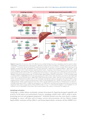
                Figure 2. Mechanisms of recurrence after HCC ablation involve multiple biological processes. Autophagy activation supports cellular
                homeostasis [24] , promoting invasion and metastasis, while EMT enhances tumor cell plasticity, fostering invasiveness and stress
                       [8]
                resistance . The hypoxic microenvironment from heat-induced damage and vascular occlusion activates HIF-1α signaling, driving
                                                 [35]                                      [3]      [41]
                metabolic reprogramming, EMT, and angiogenesis  . Immune suppression is intensified via upregulated MDSCs  and TAMs  , which
                inhibit effective anti-tumor immune responses and foster tumor growth. Collectively, these mechanisms allow residual tumor cells to
                survive, proliferate, and metastasize. Software: Adobe Illustrator (version 27.0; Adobe Inc.). HCC: Hepatocellular carcinoma; EMT:
                epithelial-mesenchymal transition; EMT-TFs: epithelial-mesenchymal transition-related transcription factors; Akt: protein kinase B; Wnt:
                Wingless/Integrated signaling pathway; ERK: extracellular signal-regulated kinase; SQSTM1: sequestosome 1; mTOR: mechanistic target
                of rapamycin; PI3K: phosphatidylinositol 3-kinase; p62: sequestosome 1/p62 protein; LC3B: microtubule-associated protein 1 light chain
                3 beta; HIF-1α: hypoxia-inducible factor 1 alpha; BNIP3: BCL2 interacting protein 3; SNAIL: Snail family transcriptional repressor 1;
                TWIST: Twist family bHLH transcription factor; SLUG: Snail family transcriptional repressor 2; β-catenin: beta-catenin; MMP-2: matrix
                metalloproteinase-2; MMP-9: matrix metalloproteinase-9; METTL1: methyltransferase-like 1; WDR4: WD repeat domain 4; m7G: N7-
                methylguanosine; TGF-β: transforming growth factor beta; VEGF: vascular endothelial growth factor; O-GlcNAcylation: O-linked β-N-
                acetylglucosaminylation; MDSC: myeloid-derived suppressor cell; TAM: tumor-associated macrophage; LAPosome: LC3-associated
                phagosome; ROS: reactive oxygen species; NOX2: NADPH oxidase 2; CCL7: C-C motif chemokine ligand 7; IL-10: interleukin-10; IL-4:
                interleukin-4; IL-12b: interleukin-12 subunit beta; IFNγ: interferon gamma; CTL: cytotoxic T lymphocyte; DC: dendritic cell.

               Autophagy activation
               Autophagy, a cellular defense mechanism, sustains homeostasis by degrading damaged organelles and
               proteins. In the tumor microenvironment, however, autophagy enables tumor cells to adapt to stress,
                                                         [24]
               promoting survival and malignant progression . Sublethal thermal stress has been shown to induce
               autophagy in cancer cell lines such as human cervical adenocarcinoma cell line (HeLa), human
               hepatocellular carcinoma cell line (Huh-7), and human hepatocellular carcinoma cell line (SMMC7721),

