
Page 83 - Read Online
P. 83
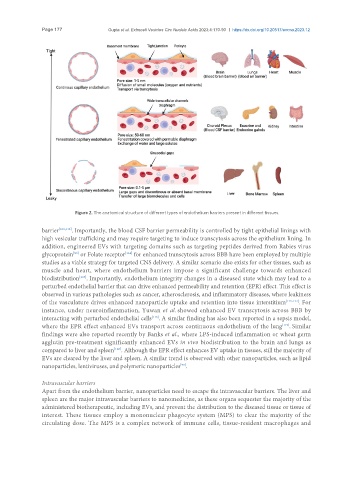
Page 177                                   Gupta et al. Extracell Vesicles Circ Nucleic Acids 2023;4:170-90  https://dx.doi.org/10.20517/evcna.2023.12


                          Figure 2. The anatomical structure of different types of endothelium barriers present in different tissues.


               barrier [109,113] . Importantly, the blood CSF barrier permeability is controlled by tight epithelial linings with
               high vesicular trafficking and may require targeting to induce transcytosis across the epithelium lining. In
               addition, engineered EVs with targeting domains such as targeting peptides derived from Rabies virus
                                           [114]
               glycoprotein  or Folate receptor  for enhanced transcytosis across BBB have been employed by multiple
                          [93]
               studies as a viable strategy for targeted CNS delivery. A similar scenario also exists for other tissues, such as
               muscle and heart, where endothelium barriers impose a significant challenge towards enhanced
                            [107]
               biodistribution . Importantly, endothelium integrity changes in a diseased state which may lead to a
               perturbed endothelial barrier that can drive enhanced permeability and retention (EPR) effect. This effect is
               observed in various pathologies such as cancer, atherosclerosis, and inflammatory diseases, where leakiness
               of the vasculature drives enhanced nanoparticle uptake and retention into tissue interstitium [115-117] . For
               instance, under neuroinflammation, Yuwan et al. showed enhanced EV transcytosis across BBB by
               interacting with perturbed endothelial cells . A similar finding has also been reported in a sepsis model,
                                                    [118]
               where the EPR effect enhanced EVs transport across continuous endothelium of the lung . Similar
                                                                                                [119]
               findings were also reported recently by Banks et al., where LPS-induced inflammation or wheat germ
               agglutin pre-treatment significantly enhanced EVs in vivo biodistribution to the brain and lungs as
                                       [120]
               compared to liver and spleen . Although the EPR effect enhances EV uptake in tissues, still the majority of
               EVs are cleared by the liver and spleen. A similar trend is observed with other nanoparticles, such as lipid
               nanoparticles, lentiviruses, and polymeric nanoparticles .
                                                              [99]

               Intravascular barriers
               Apart from the endothelium barrier, nanoparticles need to escape the intravascular barriers. The liver and
               spleen are the major intravascular barriers to nanomedicine, as these organs sequester the majority of the
               administered biotherapeutic, including EVs, and prevent the distribution to the diseased tissue or tissue of
               interest. These tissues employ a mononuclear phagocyte system (MPS) to clear the majority of the
               circulating dose. The MPS is a complex network of immune cells, tissue-resident macrophages and