
Page 82 - Read Online
P. 82
Gupta et al. Extracell Vesicles Circ Nucleic Acids 2023;4:170-90 https://dx.doi.org/10.20517/evcna.2023.12 Page 176
based on the target cell type in that specific tissue, as phagocytic cells degrade EV-associated cargo much
[80]
faster as compared to epithelial cells in a given tissue . Notably, few studies have been carried out on the in
vivo biodistribution of EVs at the cellular level. However, the general view is that Kupfer cells in the liver
phagocytose the most injected EVs [87,100-102] . A similar trend has also been observed in the spleen, where EVs
are taken up primarily by splenic macrophages . Overall liver and spleen are the major driver of EV
[102]
uptake in vivo and limits the distribution of EVs to other tissues. Notably, the liver and spleen account for
one of the multiple barriers EVs experience in vivo. Therefore, for achieving targeted delivery to a specific
tissue, EVs have to bypass four major biological barriers similar to the majority of other nanoparticle-based
drug delivery systems. These four biological barriers are as follows:
Endothelium barrier
One of the initial barriers exogenous EVs encounter upon systemic delivery is the blood-endothelium
barrier [103,104] . To achieve tissue-specific distribution, EVs need to cross the endothelial monolayer lining the
blood vessels of the tissue of interest. The permeability of these endothelial junctions is highly tissue-specific
and can have a drastic variation in adsorption efficiencies depending on the type. The endothelium can
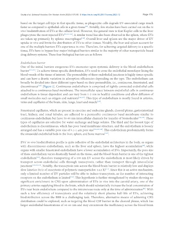
broadly be divided into three different types based on their permeability, i.e., continuous, fenestrated, and
discontinuous [Figure 2]. Continuous endothelium is comprised of tightly connected endothelial cells
[105]
attached to a continuous basal membrane. The intercellular space between endothelial cells in continuous
endothelium is tissue-dependent and can vary from 1-3 nm in healthy conditions and allow diffusion of
small molecules such as oxygen and nutrients [106,107] . This type of endothelium is mostly found in arteries,
veins and capillaries of the brain, skin, lungs, heart and muscle .
[107]
Fenestrated capillaries, which are present in exocrine and endocrine glands, choroid plexus, gastrointestinal
tract, kidney, and renal tubules, are adhered to a permeable continuous basal membrane similar to
continuous endothelium but have 50-60 nm intercellular channels for transfer of biomolecules [106,107] . These
types of capillaries are selective for water exchange and large solutes. The third and the loosest type of
endothelium is discontinuous, which has poor basal membrane structure, and the endothelium is loosely
arranged and has a variable pore size of 0.1-1 µm pore size [103,107,108] . This endothelium predominately forms
[107]
the sinusoidal endothelial beds in the liver, spleen, and bone marrow .
EVs’ in vivo biodistribution profile is quite reflective of the endothelial architecture in the body, as organs
[80]
with discontinuous endothelium, such as the liver and spleen, have the highest accumulation , while
organs with smaller fenestrated endothelium have a lower accumulation of EVs. Importantly, the pore size
of these endothelium varies drastically based on the tissue, and the blood-brain barrier is one of the tightest
endothelium , therefore transporting of a 100 nm EV across the endothelium is most likely driven by
[109]
transport across endothelial cells through transcytosis, rather than transport through intercellular
junctions [104,106,110] . Notably, the transcytosis rate across the blood-brain barrier is relatively low and displays
[111]
slow kinetics (t1/2 of exocytosis of polymeric nanoparticles: 14.2 h) . Since this is an active mechanism,
only a limited number of EV particles will be able to induce transcytosis, as the number of interacting
receptors on the endothelium is limited . This hypothesis is further strengthened by studies showing no
[112]
significant enrichment in CNS upon administration of EVs in vivo into the carotid artery, one of the
primary arteries supplying blood to the brain, which should substantially increase the local concentration of
EVs near brain endothelium compared to the intravenous route only at the time of administration . With
[80]
such a low efficiency of transcytosis and the relatively short plasma half-life of EVs, achieving
biodistribution across the BBB is a challenging task. Therefore, alternative means of achieving CNS
distribution could be explored, such as targeting the blood CSF barrier in the choroid plexus, which has
larger endothelial fenestrations of 50-60 nm and may circumvent the inefficiency across the blood-brain