
Page 80 - Read Online
P. 80
Gupta et al. Extracell Vesicles Circ Nucleic Acids 2023;4:170-90 https://dx.doi.org/10.20517/evcna.2023.12 Page 174
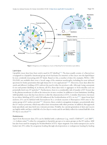
Figure 1. Exogenous and endogenous means of labelling EVs for biomedical applications.
Lipid dyes
Lipophilic tracer dyes have been widely used for EV labelling [7,76] . The dyes usually consist of a fluorophore
conjugated to a lipophilic functional group which facilitates the insertion of the tracer into the lipid bilayer
by non-covalently interacting with EV lipids. Based on this, a number of dyes (for example, PKH67, DiR/
DiL/DiD) are available that cover a broad range of the emission wavelengths, including the near-infrared
spectrum for better penetration through tissues for in vivo applications [7,76,77] . Moreover, these dyes allow for
quick and efficient labelling of EVs without the need to alter the producer cells. Although being convenient
to use and permit labelling of, in theory, all EVs, these dyes tend to aggregate or form micelles and can
[78]
potentially label non-EV particles . Furthermore, there is a considerable risk of transfer of EV-bound dye
to the plasma membrane of cells as the interaction is non-covalent. In addition to these limitations, labelling
with lipophilic tracer dye has been shown to alter the characteristics of EVs. A similar observation was made
by us where labelling of EVs with DiR influenced the biodistribution of EVs in vivo [79,80] . Apart from lipid
anchors, EVs can be labelled with fluorophores by a covalent reaction of fluorophore NHS ester to the
amine group of EV surface proteins [81,82] . However, these covalent conjugation strategies can potentially alter
the EV surface proteome, which may affect their interactions with other proteins. In addition, this approach
lacks specificity and may label non-vesicular proteins. Notably, the current generation of dyes and tracers
are highly stable and have a half-life of a few days to weeks and do not likely reflect the natural half-life of an
EV that is very short [83,84] .
Radiotracers
[85]
Apart from fluorescent dyes, EVs can be labelled with a radiotracer (e.g., 99mTc-HMPAO , 125I-IBB ,
[86]
111-Indium-oxine ) either by conjugation to lipophilic groups or to amine groups on the EV surface. MRI
[87]
has also been used for imaging the biodistribution of EVs. Super magnetic iron oxide nanoparticles can be
loaded into EVs either by exogenous loading through electroporation or endogenous loading by feeding