
Page 109 - Read Online
P. 109
Tutanov et al. Extracell Vesicles Circ Nucleic Acids 2023;4:195-217 https://dx.doi.org/10.20517/evcna.2023.17 Page 207
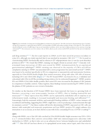
Figure 6. Two methods of sorting apical and basolateral proteins into the same EV. The left side depicts the progressive loss of polarity
during CRC progression, leading to apical (DPEP1) and basolateral (EGFR) cell-surface proteins being in the same EV. The right side
depicts different routes of trafficking of apical and basolateral proteins, which can lead to their presence in the common recycling
[104]
endosome . This endosome can then mature into an MVB, which would contain both DPEP1 and EGFR in the same intraluminal
vesicle.
and drug resistance [109-112] . The few scant reports on DPEP1 in EVs have noted its presence in kidney or
urine EVs, although one study reported that it was enriched in metastatic CRC EVs [113-115] . We focused on
characterizing DPEP1 biochemically and in relation to EV subpopulations since it was the most abundant
protein in sEVs . We found that DPEP1 staining was largely absent in normal colon . Clinically well-
[13]
[13]
annotated tissue microarrays of CRCs were scored for DPEP1 immunoreactivity by an experienced
[13]
gastrointestinal pathologist . DPEP1 staining was observed in the majority of CRCs and diffuse
cytoplasmic staining for DPEP1 was associated with a worse progression-free and overall survival . DPEP1
[13]
was found to be enriched specifically in EVs in comparison to nanoparticles and nonvesicular fractions,
especially in CD81/EGFR double-bright, flow-sorted exosomes, along with other GPI-APs of interest,
including CD73 and CEACAM5 [Figure 5] . The EV-bound DPEP1 was found to be α2, 6-sialylated and
[13]
[13]
colocalized with CD63 in MVBs, providing strong evidence for its endosomal biogenesis . DPEP1-positive
EVs and DPEP1/CEACAM5 double-positive, flow-sorted EVs were increased in the plasma of CRC patients
in comparison to healthy controls. Taken together, these findings warrant monitoring EV DPEP1 levels in
the plasma of CRC patients as a non-invasive biomarker.
No studies on the function of EV-bound DPEP1 have been reported, but there is a growing body of
literature concerning a new nonenzymatic function of DPEP1, that is, binding neutrophils and
monocytes [116,117] . These reports show that DPEP1 can be expressed by endothelial cells in response to an
inflammatory insult, resulting in increased neutrophil and monocyte binding and infiltration [116,117] .
Interestingly, both the enzymatic inhibitor and neutrophil-binding inhibitor of DPEP1 reduce immune cell
recruitment and binding, suggesting that DPEP1 might have a role in producing a chemoattractant through
its enzymatic activity . Our future studies will involve determining if DPEP1 expressed on CRC cells can
[117]
interact with neutrophil and monocytic populations and how DPEP1-positive EVs might enhance this
interaction or act as decoys, as we have shown occurs with ACE2-containing EVs in a COVID-19 setting .
[118]
CD73
Along with DPEP1, one of the GPI-APs enriched in CD81/EGFR double-bright exosomes was CD73. CD73
is a 5’-ectonucleotidase that converts extracellular AMP into immunosuppressive adenosine with
similarities to DPEP1 in that it also acts as an adhesion factor between lymphocytes and endothelial
cells [119,120] . This GPI-AP has been shown to be upregulated across many cancer types, and it has roles in