
Page 221 - Read Online
P. 221
Kautsar et al. Energy Mater. 2025, 5, 500129 https://dx.doi.org/10.20517/energymater.2025.26 Page 5 of 14
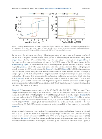
Figure 1. (A) Magnetization curves of HP and HD magnets, showing the transition from isotropic to anisotropic magnetic properties;
(B) Magnetization curves of HD magnets after Dy-Nd-Cu, Nd-Cu, and Pr-Cu GBDP, illustrating retained anisotropy with varying
coercivity enhancements. HP: Hot-pressed; HD: hot-deformed; GBDP: grain boundary diffusion process.
To investigate the microstructural changes following processing, microstructural analyses were conducted
on the studied magnets. Due to differences in grain size, the HP magnet was examined using TEM
[Figure 2A], while the HD and GBDP HD magnets were observed using SEM [Figure 2B-E]. A
backscattered electron scanning electron microscopy (BSE-SEM) image of the HP magnet is provided in
Supplementary Figure 2 to illustrate the difficulty of observing its fine-grained structure using SEM. The HP
magnet [Figure 2A] exhibits fine, equiaxed grains with sizes less than 100 nm. These isotropic grains
contribute to the low M /M ratio observed in Figure 1A. After HD [Figure 2B], the Nd Fe B grains evolve
s
2
14
r
into well-aligned, platelet-like grains with sizes exceeding 200 nm in lateral direction. Note that the brightly
imaged regions in BSE-SEM images indicate the presence of a Nd-rich phase existing in the grain boundary
region of the HD sample. This microstructural transformation explains the increase in the M /M ratio after
s
r
HD [Figure 1A], which shifts the magnet’s characteristics from isotropic to anisotropic. The observed
anisotropic grains after HD is consistent with the previous reports . However, unlike in , platelet-shaped
[38]
[38]
grains were not observed in the HP magnet in this study, likely due to the short duration (less than five
minutes) of the hot pressing process.
Figure 2C-E illustrates the microstructures of the RE-Cu (RE = Dy-Nd, Nd, Pr) GBDP magnets. These
images reveal a significant change in the thickness of RE-rich IGP following RE-Cu GBDP, evident from the
increased areal fraction of the bright phase in BSE-SEM images, from 6% in the HD magnet [Figure 2B] to
20%, 19%, and 28% after Dy-Nd-Cu, Nd-Cu, and Pr-Cu GBDP, respectively [Figure 2C-E]. The formation
of a thick RE-rich IGP, which magnetically isolates Nd Fe B grains, is known to enhance coercivity in the
14
2
GBDP magnets [39-41] . In addition, grain misorientations and the increased volume fraction of the IGP
observed in the RE-Cu GBDP magnets likely account for the reduced M in these magnets, as presented in
r
Figure 1B.
We investigated the microstructure and the distribution of constituent and diffused elements in HD and
RE-Cu (RE = Dy-Nd, Nd, Pr) GBDP magnets using high-angle annular dark field (HAADF)-scanning
transmission electron microscopy (STEM) and STEM-energy dispersive X-ray spectroscopy (EDS)
techniques [Figure 3A-D]. Our observations revealed an increase in thickness and segregation of RE-Cu
elements within the IGP of the GBDP magnets. Additionally, we identified the formation of (Nd,Dy) Fe B
14
2