
Page 45 - Read Online
P. 45
Page 724 Bijnsdorp et al. Cancer Drug Resist 2021;4:719-27 https://dx.doi.org/10.20517/cdr.2021.21
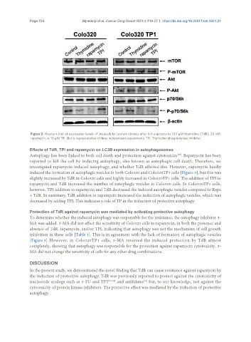
Figure 2. Western blot of expression levels of intracellular protein kinases after 6 h exposure to 100 µM thymidine (TdR), 20 nM
rapamycin, or 10 µM TPI. Blot is representative of three independent experiments. TPI: Thymidine phosphorylase inhibitor.
Effects of TdR, TPI and rapamycin on LC3B expression in autophagosomes
[23]
Autophagy has been linked to both cell death and protection against cytotoxicity . Rapamycin has been
reported to kill the cell by inducing autophagy, also known as autophagic cell death. Therefore, we
investigated rapamycin induced autophagy, and whether TdR affected this. However, rapamycin hardly
induced the formation of autophagic vesicles in both Colo320 and Colo320TP1 cells [Figure 3], but this was
slightly increased by TdR in Colo320 cells and highly increased in Colo320TP1 cells. The addition of TPI to
rapamycin and TdR increased the number of autophagic vesicles in Colo320 cells. In Colo320TP1 cells,
however, TPI addition to rapamycin and TdR decreased the induced autophagic vesicles compared to Rapa
+ TdR. In summary, TdR addition to rapamycin increased the induction of autophagic vesicles, which was
decreased by adding TPI. This indicates a role of TP in the induction of protective autophagy.
Protection of TdR against rapamycin was mediated by activating protective autophagy
To determine whether the induced autophagy was responsible for the resistance, the autophagy inhibitor 3-
MA was added. 3-MA did not affect the sensitivity of Colo320 cells to rapamycin, in both the presence and
absence of TdR, rapamycin, and/or TPI, indicating that autophagy was not the mechanism of cell growth
inhibition in these cells [Table 1]. This is in agreement with the lack of formation of autophagic vesicles
[Figure 3]. However, in Colo320TP1 cells, 3-MA reversed the induced protection by TdR almost
completely, showing that autophagy was responsible for the protection against rapamycin cytotoxicity. 3-
MA did not change the sensitivity of cells for any other drug combinations.
DISCUSSION
In the present study, we demonstrated the novel finding that TdR can cause resistance against rapamycin by
the induction of protective autophagy. TdR was previously reported to protect against the cytotoxicity of
[35]
nucleoside analogs such as 5-FU and TFT [33,34] and antifolates but, to our knowledge, not against the
cytotoxicity of protein kinase inhibitors. The protective effect was mediated by the induction of protective
autophagy.