
Page 62 - Read Online
P. 62
Gurska et al. Cancer Drug Resist 2023;6:674-87 https://dx.doi.org/10.20517/cdr.2023.39 Page 678
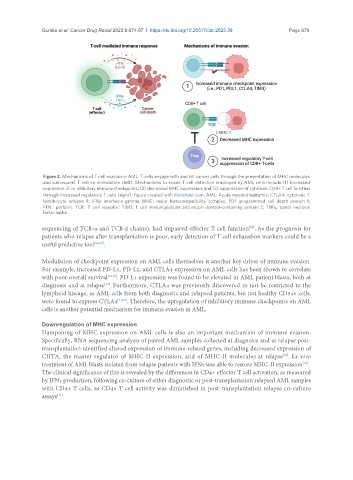
Figure 2. Mechanisms of T cell evasion in AML. T cells engage with and kill cancer cells through the presentation of MHC molecules
and subsequent T cell co-stimulation (left). Mechanisms to evade T cell detection employed by AML cells include (1) increased
expression of co-inhibitory immune checkpoints; (2) decreased MHC expression; and (3) suppression of cytotoxic CD8+ T cell function
through increased regulatory T cells (right). Figure created with Biorender.com. AML: Acute myeloid leukemia; CTLA4: cytotoxic T
lymphocyte antigen 4; IFNγ: interferon gamma; MHC: major histocompatibility complex; PD1: programmed cell death protein 1;
PFN: perforin; TCR: T cell receptor; TIM3: T cell immunoglobulin and mucin domain-containing protein 3; TNFα: tumor necrosis
factor alpha.
sequencing of TCR-α and TCR-β chains), had impaired effector T cell function . As the prognosis for
[27]
patients who relapse after transplantation is poor, early detection of T cell exhaustion markers could be a
useful predictive tool [26,27] .
Modulation of checkpoint expression on AML cells themselves is another key driver of immune evasion.
For example, increased PD-L1, PD-L2, and CTLA4 expression on AML cells has been shown to correlate
with poor overall survival [28,29] . PD-L1 expression was found to be elevated in AML patient blasts, both at
diagnosis and at relapse . Furthermore, CTLA4 was previously discovered to not be restricted to the
[30]
lymphoid lineage, as AML cells from both diagnostic and relapsed patients, but not healthy CD34+ cells,
were found to express CTLA4 [31,32] . Therefore, the upregulation of inhibitory immune checkpoints on AML
cells is another potential mechanism for immune evasion in AML.
Downregulation of MHC expression
Dampening of MHC expression on AML cells is also an important mechanism of immune evasion.
Specifically, RNA sequencing analysis of paired AML samples collected at diagnosis and at relapse post-
transplantation identified altered expression of immune-related genes, including decreased expression of
CIITA, the master regulator of MHC-II expression, and of MHC-II molecules at relapse . Ex vivo
[33]
[33]
treatment of AML blasts isolated from relapse patients with IFNγ was able to restore MHC-II expression .
The clinical significance of this is revealed by the differences in CD4+ effector T cell activation, as measured
by IFNγ production, following co-culture of either diagnostic or post-transplantation relapsed AML samples
with CD4+ T cells, as CD4+ T cell activity was diminished in post-transplantation relapse co-culture
assays .
[33]