
Page 61 - Read Online
P. 61
Page 677 Gurska et al. Cancer Drug Resist 2023;6:674-87 https://dx.doi.org/10.20517/cdr.2023.39
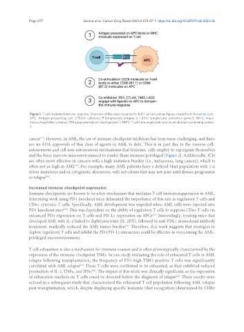
Figure 1. T cell-mediated immune response. Overview of the steps required for full T cell activation. Figure created with Biorender.com.
APC: Antigen-presenting cell; CTLA4: cytotoxic T lymphocyte antigen 4; LAG3: lymphocyte activation gene-3; MHC: major
histocompatibility complex; PD1: programmed cell death protein 1; TIM3: T cell immunoglobulin and mucin domain-containing protein
3.
[22]
cancer . However, in AML, the use of immune checkpoint inhibitors has been more challenging, and there
are no FDA approvals of this class of agents in AML to date. This is in part due to the various cell-
autonomous and cell non-autonomous mechanisms that leukemic cells employ to reprogram themselves
and the bone marrow microenvironment to render them immune privileged [Figure 2]. Additionally, ICIs
are often most effective in cancers with a high mutation burden (i.e., melanoma, lung cancer), which is
often not as high in AML . For example, many AML patients have a defined blast population with 1-2
[23]
driver mutations and/or cytogenetic alterations, with sub-clones that may not arise until disease progression
or relapse .
[24]
Increased immune checkpoint expression
Immune checkpoints are known to be a key mechanism that mediates T cell immunosuppression in AML.
Interesting work using PD1 knockout mice delineated the importance of this axis in regulatory T cells and
CD8+ cytotoxic T cells. Specifically, AML development was impeded when AML cells were injected into
PD1 knockout mice . This was dependent on the ability of regulatory T cells to suppress CD8+ T cells via
[25]
enhanced PD1 expression on T cells and PD-L1 expression on APCs . Interestingly, treating mice that
[25]
developed AML with IL-2 linked to diphtheria toxin (IL-2DT), followed by anti-PDL1 monoclonal antibody
treatment, markedly reduced the AML tumor burden . Therefore, this work suggests that strategies to
[25]
deplete regulatory T cells and inhibit the PD1/PD-L1 interaction could be effective in overcoming the AML-
privileged microenvironment.
T cell exhaustion is also a mechanism for immune evasion and is often phenotypically characterized by the
expression of the immune checkpoint TIM3. In one study evaluating the role of exhausted T cells in AML
relapse following transplantation, the frequency of PD1-high TIM3-positive T cells was significantly
correlated with AML relapse . These T cells were confirmed to be exhausted, as they exhibited reduced
[26]
[26]
production of IL-2, TNFα, and IFNγ . The impact of this study was clinically significant, as the expression
of exhaustion markers on T cells could be detected before the diagnosis of relapse . These results were
[26]
echoed in a subsequent study that characterized the exhausted T cell population following AML relapse
post-transplantation, which, despite displaying specific leukemic blast recognition (determined by CDR3