
Page 102 - Read Online
P. 102
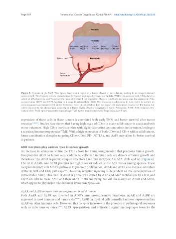
Page 757                                            Remley et al. Cancer Drug Resist 2023;6:748-67  https://dx.doi.org/10.20517/cdr.2023.63


                Figure 3. Hypoxia in the TME. This figure illustrates a region of a tumor devoid of vasculature, leading to an oxygen-starved
                environment. This hypoxic zone is characterized by low pH and a predominance of lactate. Within this environment, TAMs tend to
                adopt an M2 phenotype, and Tregs become the predominant T cell population. Hypoxic conditions also encourage the expression of the
                ectoenzymes CD39 and CD73, leading to a surge in extracellular ADO. This increase in adenosine, in turn, helps to sustain an
                immunosuppressive environment within the tumor. Note: this illustration does not depict the anatomical structure of the tumor, but
                rather represents the phenomena occurring at different levels of tumor oxygenation. ADO: Adenosine; A2BR: A2B receptors; NK:
                natural killer; TAM: tumor-associated macrophage; TME: tumor microenvironment; Tregs: regulatory T cells.


               expression of these cells in these tumors is correlated with early TNM and better survival after tumor
               resection [132,133] . Studies have shown that having high levels of CD73 in many solid tumors is associated with
               worse outcomes. High CD73 levels correlate with higher adenosine concentrations in the tumor, leading to
               a sustained immunosuppressive TME. With a high expression of both CD39 and CD73 within solid tumors,
               future combination therapies targeting CD39/CD73, PD-1/CTLA4, and A2BR may allow for better survival
               in patients.

               ADO receptors play various roles in cancer growth
               An increase in adenosine within the TME allows for immunosuppression that promotes tumor growth.
               Receptors for ADO on tumor cells, endothelial cells, and immune cells are drivers of tumor growth and
               metastasis. The ADO G-protein coupled receptors have four subtypes: A1, A2A, A2B, and A3 [Figure 4].
               The A1R, A2AR, and A2BR proteins are highly conserved, while the A3R varies among species. These
               receptors interact with MAPK pathways to promote proliferation. A2AR and A2BR also increase activation
               of the mTOR and ERK pathways . However, receptor signaling is dependent on the concentration of
                                            [134]
               extracellular ADO. This level of ADO is primarily dictated by ATP and ADP metabolism by CD39 and
               CD73 on cells to make AMP and then ADO. In the following, we will focus only on A2AR and A2BR,
               which appear to play major roles in tumor immunosuppression.


               A2AR and A2BR increase immunosuppression in solid tumors
               Both A2AR and A2BR are involved in ADO’s immunosuppressive functions. A2AR and A2BR are
               expressed in most immune and tumor cells [59,135] . A2BR on myeloid cells normally has lower expression than
               A2AR on other immune cells. However, this receptor increases in the presence of pathological responses
               such as infections or cancer . A2BR upregulation and activation signal macrophages towards the
                                         [136]