
Page 98 - Read Online
P. 98
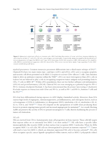
Page 753                                           Remley et al. Cancer Drug Resist 2023;6:748-67  https://dx.doi.org/10.20517/cdr.2023.63


                Figure 1. Adenosine’s pleiotropic effects on immune cells. ADO facilitates the evasion of tumor cells from immune detection by
                restricting the activity of T cells, DCs, NK cells, macrophages, and neutrophils. Concurrently, adenosine amplifies the functionality of
                immunosuppressive cell types like MDSCs and Tregs. ADO: Adenosine; A2AR: A2A receptors; A2BR: A2B receptors; DCs: dendritic
                cells; MDSCs: myeloid-derived suppressor cells; NK: natural killer; TNFα: tumor necrosis factor-alpha; Tregs: regulatory T cells; VEGF:
                vascular endothelial growth factor.


               myeloid precursors. Common monocyte precursors differentiate into a third major subtype of DCs .
                                                                                                       [80]
               Classical DCs have two major states: type 1 and type 2 (cDC1 and cDC2). cDC1 acts to recognize apoptotic
               and necrotic cell debris presented on its MHC-I receptors to activate CD8+ effector T cells. Their function
               helps to drive an antitumor response within the TME . cDC2 are more heterogenous than cDC1 cells in
                                                             [75]
               tumors but are believed to play a role in recognizing exogenous tumor antigens and presenting them to
               CD4+ T cells on MHC-II . Within cDC2 populations, there are two further subtypes: anti-inflammatory
                                     [81]
               (cDC2A) and proinflammatory (cDC2B). Classical DCs are now emerging as a potential target for PD-1/
               PD-L1 immune checkpoint blockade. It has been demonstrated that the proper functioning of checkpoint
               blockade requires cis interactions with CD80 and PD-L1, as well as PD-1 and PD-L1, between T cells and
               the DCs [82,83] .

               DCs that have differentiated during exposure to ADO display diminished activity. Moreover, these DCs
               express high levels of angiogenic, immunosuppressive, proinflammatory, and tolerogenic factors, such as
               cyclooxygenase-2 (COX-2), indoleamine 2,3-dioxygenase (IDO), interleukin-6 (IL-6), interleukin-8 (IL-8),
               IL-10, TGF-β, and VEGF [84,85] . These DCs depend on the upregulation of A2BR when producing these
               factors to promote ongoing tumor growth and increased angiogenesis for metastasis . As a result, blocking
                                                                                      [85]
               A2BR can preserve the activity of DCs to present neoantigens to T cells, thereby facilitating the process of
               tumor cell destruction.


               NK cells
               NKs are derived from CD34+ hematopoietic stem cell progenitors in bone marrow. These cells kill targets
               that express either no or extremely low MHC-I on their surface [86,87] . NK cells have a specific killer
               immunoglobulin-like receptor (KIR) on their surface that recognizes MHC-I molecules. When the KIRs
               recognize MHC-I in self-cells, NK cells are downregulated to prevent an immune response [86,88,89] . Tumor
               cells tend to have low MHC-I, which can stimulate suppressed NK cells to become activated . NK cells
                                                                                                [90]
               often recognize specific cancer ligands upregulated within tumors, such as MHC-I polypeptide-related