
Page 81 - Read Online
P. 81
Choi et al. Cancer Drug Resist. 2026;9:12 Page 7 of 20
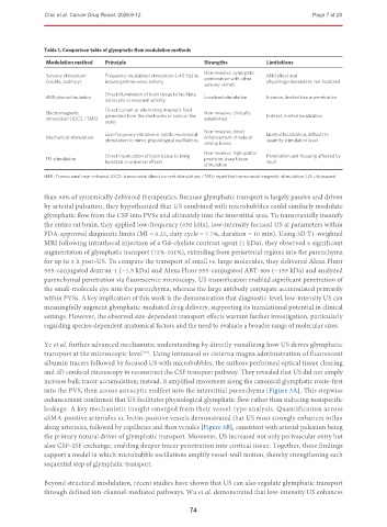
Table 1. Comparison table of glymphatic flow modulation methods
Modulation method Principle Strengths Limitations
Non-invasive, synergistic
Sensory stimulation Frequency-modulated stimulation (~40 Hz) to Mild effect and
(visible, auditory) induce gamma-wave activity combination with other physiology-dependent, not localized
sensory stimuli
Direct illumination of brain tissue to facilitate
tNIR photostimulation Localized stimulation Invasive, limited tissue penetration
astrocytic or neuronal activity
Direct current or alternating magnetic field
Electromagnetic Non-invasive, clinically
stimulation (tDCS, rTMS) generated from the electrodes or coils on the established Indirect, limited localization
scalp
Non-invasive, direct
Low-frequency vibration or subtle mechanical Limited localization, difficult to
Mechanical stimulation enhancement of natural
stimulation to mimic physiological oscillations quantify stimulation level
driving forces
Non-invasive, high spatial
Direct insonication of brain tissue to bring Penetration and focusing affected by
US stimulation precision, deep tissue
localized or universal effects skull
stimulation
tNIR: Transcranial near-infrared; tDCS: transcranial direct current stimulation; rTMS: repetitive transcranial magnetic stimulation; US: ultrasound.
than 98% of systemically delivered therapeutics. Because glymphatic transport is largely passive and driven
by arterial pulsation, they hypothesized that US combined with microbubbles could similarly modulate
glymphatic flow from the CSF into PVSs and ultimately into the interstitial area. To transcranially insonify
the entire rat brain, they applied low-frequency (650 kHz), low-intensity focused US at parameters within
FDA-approved diagnostic limits (MI = 0.25, duty cycle = 7.7%, duration = 10 min). Using 3D T1-weighted
MRI following intrathecal injection of a Gd-chelate contrast agent (1 kDa), they observed a significant
augmentation of glymphatic transport (72%-101%), extending from periarterial regions into the parenchyma
for up to 3 h post-US. To compare the transport of small vs. large molecules, they delivered Alexa Fluor
555-conjugated dextran-1 (~1.5 kDa) and Alexa Fluor 555-conjugated ABT-806 (~155 kDa) and analyzed
parenchymal penetration via fluorescence microscopy. US insonification enabled significant penetration of
the small-molecule dye into the parenchyma, whereas the large antibody conjugate accumulated primarily
within PVSs. A key implication of this work is the demonstration that diagnostic-level, low-intensity US can
meaningfully augment glymphatic-mediated drug delivery, supporting its translational potential in clinical
settings. However, the observed size-dependent transport effects warrant further investigation, particularly
regarding species-dependent anatomical factors and the need to evaluate a broader range of molecular sizes.
Ye et al. further advanced mechanistic understanding by directly visualizing how US drives glymphatic
transport at the microscopic level . Using intranasal or cisterna magna administration of fluorescent
[63]
albumin tracers followed by focused US with microbubbles, the authors performed optical tissue clearing
and 3D confocal microscopy to reconstruct the CSF transport pathway. They revealed that US did not simply
increase bulk tracer accumulation; instead, it amplified movement along the canonical glymphatic route–first
into the PVS, then across astrocytic endfeet into the interstitial parenchyma [Figure 3A]. This stepwise
enhancement confirmed that US facilitates physiological glymphatic flow rather than inducing nonspecific
leakage. A key mechanistic insight emerged from their vessel-type analysis. Quantification across
αSMA-positive arterioles vs. lectin-positive vessels demonstrated that US most strongly enhances influx
along arterioles, followed by capillaries and then venules [Figure 3B], consistent with arterial pulsation being
the primary natural driver of glymphatic transport. Moreover, US increased not only perivascular entry but
also CSF–ISF exchange, enabling deeper tracer penetration into cortical tissue. Together, these findings
support a model in which microbubble oscillations amplify vessel-wall motion, thereby strengthening each
sequential step of glymphatic transport.
Beyond structural modulation, recent studies have shown that US can also regulate glymphatic transport
through defined ion-channel-mediated pathways. Wu et al. demonstrated that low-intensity US enhances
74