
Page 136 - Read Online
P. 136
Wang et al. Cancer Drug Resist. 2026;9:8 Page 3 of 18
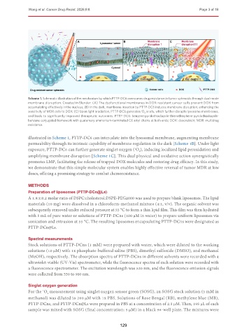
Scheme 1. Schematic illustration of the mechanism by which PTTP-DC6 overcomes drug resistance in tumor spheroids through dual-mode
membrane disruption. Created in Blender. (A) The dysfunctional membranes in DOX-resistant cancer cells prevent DOX from
accumulating effectively in the nucleus; (B) In the dark, membrane insertion by PTTP-DC6 induces membrane disruption, enhancing the
sensitivity of MDR cells to DOX; (C) Upon light irradiation, PTTP-DC6 generates O 2 in situ, which further disrupts lysosome membranes,
1
and leads to significantly improved therapeutic outcomes. PTTP-DC6: benzene-pyridothiadiazole-thienothiophene-pyridothiadiazole-
benzene conjugated framework with quaternary ammonium-terminated C6 alkyl chains at both ends; DOX: doxorubicin; MDR: multidrug
resistance.
illustrated in Scheme 1, PTTP-DC6 can intercalate into the lysosomal membrane, augmenting membrane
permeability through its intrinsic capability of membrane regulation in the dark [Scheme 1B]. Under light
exposure, PTTP-DC6 can further generate singlet oxygen ( O ), inducing localized lipid peroxidation and
1
2
amplifying membrane disruption [Scheme 1C]. This dual physical and oxidative action synergistically
promotes LMP, facilitating the release of trapped DOX molecules and restoring drug efficacy. In this study,
we demonstrate that this simple molecular system enables highly effective reversal of tumor MDR at low
doses, offering a promising strategy to combat chemoresistance.
METHODS
Preparation of liposomes (PTTP-DCn@Ls)
A 1.8:1:0.2 molar ratio of DSPC:cholesterol:DSPE-PEG2000 was used to prepare blank liposomes. The lipid
materials (10 mg) were dissolved in a chloroform-methanol mixture (4:1, v/v). The organic solvent was
subsequently removed under reduced pressure at 55 °C to form a thin lipid film. This film was then hydrated
with 5 mL of pure water or solutions of PTTP-DCns (100 μM in water) to prepare uniform liposomes via
sonication and extrusion at 55 °C. The resulting liposomes encapsulating PTTP-DCns were designated as
PTTP-DCn@Ls.
Spectral measurements
Stock solutions of PTTP-DCns (1 mM) were prepared with water, which were diluted to the working
solutions (10 μM) with 1x phosphate-buffered saline (PBS), dimethyl sulfoxide (DMSO), and methanol
(MeOH), respectively. The absorption spectra of PTTP-DCns in different solvents were recorded with a
ultraviolet-visible (UV-Vis) spectrometer, while the fluorescence spectra of each solution were recorded with
a fluorescence spectrometer. The excitation wavelength was 520 nm, and the fluorescence emission signals
were collected from 550 to 900 nm.
Singlet oxygen generation
For the O measurement using singlet oxygen sensor green (SOSG), an SOSG stock solution (5 mM in
1
2
methanol) was diluted to 200 μM with 1x PBS. Solutions of Rose Bengal (RB), methylene blue (MB),
PTTP-DCns, and PTTP-DCn@Ls were prepared in PBS at a concentration of 2.5 μM. Then, 195 μL of each
sample was mixed with SOSG (final concentration: 5 μM) in a black 96-well plate. The mixtures were
129