
Page 75 - Read Online
P. 75
Johnson et al. Art Int Surg 2024;4:401-10 https://dx.doi.org/10.20517/ais.2024.40 Page 405
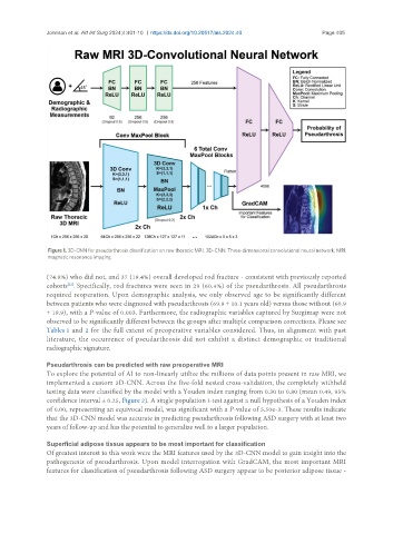
Figure 1. 3D-CNN for pseudarthrosis classification on raw thoracic MRI. 3D-CNN: Three-dimensional convolutional neural network; MRI:
magnetic resonance imaging.
(74.9%) who did not, and 37 (19.4%) overall developed rod fracture - consistent with previously reported
[22]
cohorts . Specifically, rod fractures were seen in 29 (60.4%) of the pseudarthrosis. All pseudarthrosis
required reoperation. Upon demographic analysis, we only observed age to be significantly different
between patients who were diagnosed with pseudarthrosis (69.9 ± 10.1 years old) versus those without (60.9
± 19.9), with a P-value of 0.003. Furthermore, the radiographic variables captured by Surgimap were not
observed to be significantly different between the groups after multiple comparison corrections. Please see
Tables 1 and 2 for the full extent of preoperative variables considered. Thus, in alignment with past
literature, the occurrence of pseudarthrosis did not exhibit a distinct demographic or traditional
radiographic signature.
Pseudarthrosis can be predicted with raw preoperative MRI
To explore the potential of AI to non-linearly utilize the millions of data points present in raw MRI, we
implemented a custom 3D-CNN. Across the five-fold nested cross-validation, the completely withheld
testing data were classified by the model with a Youden index ranging from 0.30 to 0.80 (mean 0.49, 95%
confidence interval ± 0.25, Figure 2). A single population t-test against a null hypothesis of a Youden index
of 0.00, representing an equivocal model, was significant with a P-value of 5.50e-3. These results indicate
that the 3D-CNN model was accurate in predicting pseudarthrosis following ASD surgery with at least two
years of follow-up and has the potential to generalize well to a larger population.
Superficial adipose tissue appears to be most important for classification
Of greatest interest to this work were the MRI features used by the 3D-CNN model to gain insight into the
pathogenesis of pseudarthrosis. Upon model interrogation with GradCAM, the most important MRI
features for classification of pseudarthrosis following ASD surgery appear to be posterior adipose tissue -