
Page 94 - Read Online
P. 94
Xu et al. Art Int Surg 2023;3:48-63 https://dx.doi.org/10.20517/ais.2022.33 Page 52
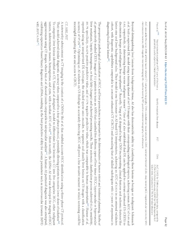
Preis et al. [48] Neural network evaluation of PET scans of the liver: a Identify metastatic liver disease PET CNN AUC: 0.905 for CNN incorporating lesion data, compared to
potentially useful adjunct in clinical interpretation 0.786 for blinded observers; 0.896 for CNN independent of
lesion data, compared to 0.796 for blinded observers
AUC: area under the curve; ANN: artificial neural network; CT: computed tomography; CNN: convolutional neural networks; CEUS: contrast-enhanced US; DL: deep learning HCC: Hepatocellular carcinoma; MVI:
microvascular invasion; MRI: magnetic resonance imaging; ML: machine learning; PET: positron emission tomography; US: ultrasound.
Beyond distinguishing liver lesions from background tissue, AI also has demonstrable utility in classifying these lesions as benign or malignant. Schmauch
et al. built a supervised DL model using a training dataset of 367 US images with their corresponding radiological reports, achieving a mean AUC of 0.93 and
[15]
0.92, respectively, in determining benign versus malignant masses . Guo et al. also established that DL can be applied to contrast-enhanced US (CEUS) to
[41]
discriminate benign and malignant liver neoplasms . Recently, Yang et al. designed a large CNN incorporating clinical features and radiomic features like
lesion size and liver background echo. Their model is one forerunner in AI-based US interpretation, achieving an AUC of 0.924 in an external validation
cohort, with diagnostic capabilities comparable to contrast-enhanced CT (CECT) and exceeding that of skilled radiologists with 15 years of experience in
diagnosing focal liver lesions .
[42]
The preoperative pathological classification of HCC and liver parenchyma is important to the determination of tumor extent and treatment planning. Streba et
al. prospectively studied CEUS images of 112 patients to train an ANN that classified five different types of liver tissue (HCC, hypervascular or hypovascular
liver metastasis, hepatic hemangioma, or focal fatty changes) and achieved promising results. Their automatic classification process achieved 93.2% sensitivity,
89.7% specificity, 94.42% positive predictive value, and 87.57% negative predictive value, which was comparable to human interpretation . Hassan et al.
[43]
reported using an unsupervised DL technique, the stacked sparse auto-encoder, to segment and classify liver lesions on US images with a classification
[44]
accuracy of 97.2% . Optimizing an AI solution on US findings in accurately detecting HCC will prove a less invasive manner in which screening could be
meaningful (negating the use and access to CECT).
CT, MRI, PET
A noteworthy advancement in CT imaging is the creation of a CNN by Shi et al. that enabled accurate HCC identification using a three-phase CT protocol.
Their model achieved similar diagnostic accuracy when compared to a four-phase protocol, potentially allowing patients to receive lower doses of radiation .
[45]
To categorize liver lesions identified on CT, Yasaka et al. designed a model to differentiate liver lesions on CT into five categories: HCC, other malignant
[46]
tumors, indeterminate masses, hemangiomas, and cysts, with a median AUC of 0.92 . Most recently, the LiSNet AI tool was developed for staging of HCC
aggressiveness using CT images, where Sun et al. showed results comparable to subspecialist analysis . A human-AI partnered diagnosis was also attempted,
[21]
combining experience-based binary diagnosis and LiSNet, resulting in the best predictive ability for certain parameters such as microvascular invasion (MVI)
with AUC 0.705 .
[21]