
Page 93 - Read Online
P. 93
Page 51 Xu et al. Art Int Surg 2023;3:48-63 https://dx.doi.org/10.20517/ais.2022.33
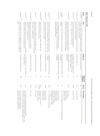
Table 1. Diagnosis of HCC
Diagnostic
Study Title Study aim AI tool Performance
technique
[38]
Liu et al. Learning to diagnose Cirrhosis with liver capsule Guided Early identification of cirrhosis US ML AUC: 0.968
ultrasound image classification
[39]
Ksiazek et al. A novel machine learning Approach for early detection of Prediction of HCC risk US ML Accuracy: 88.5%
hepatocellular carcinoma Patients
[14]
Bharti et al. Preliminary study of chronic liver classification on Classification of liver disease into four US ANN Accuracy: 96.6%
ultrasound images using an ensemble model stages (normal liver, chronic liver
disease, cirrhosis and HCC)
[40]
Brehar et al. Comparison of deep-learning and conventional machine- Differentiate HCC from cirrhotic US CNN AUC: 0.95
learning methods for the automatic recognition of the parenchyma Accuracy: 0.91
hepatocellular carcinoma areas from ultrasound Images Sensitivity: 94.4%
Specificity: 88.4%
Schmauch Diagnosis of focal liver lesions from ultrasound using deep Classification of liver lesions as benign US DL AUC: 0.93 for benign lesions, 0.92 for malignant lesions
et al. [15] learning or malignant
[41]
Guo et al. A two-stage multi-view learning framework-based Classification of liver lesions as benign CEUS ML Accuracy: 90.41%
computer-aided diagnosis of liver tumors with contrast or malignant Sensitivity: 93.56%
enhanced ultrasound images Specificity: 86.89%
Youden index: 79.44%
False positive rate: 13.11%
False negative rate: 6.44%
Yang et al. [42] Improving B-mode ultrasound diagnostic performance for Classification of liver lesions as benign US CNN AUC: 0.924 (external validation)
focal liver lesions using deep learning: A multi-center study or malignant
Streba et al. [43] Contrast-enhanced ultrasonography parameters in neural Classification of focal liver lesions US ANN Accuracy: 87.12%
network diagnosis of liver tumors Sensitivity: 93.2%
Specificity: 89.7%
[44]
Hassan et al. Diagnosis of focal liver diseases based on deep learning Classification of focal liver lesions US Auto- Accuracy: 97.2% accuracy
technique for ultrasound images encoder Sensitivity: 98%
Specificity: 95.70%
[45]
Shi et al. Deep learning assisted differentiation of hepatocellular Classification of focal liver lesions CT CNN AUC: 0.925
carcinoma from focal liver lesions: choice of four-phase and
three-phase CT imaging protocol
Yasaka et al. [46] Deep learning with convolutional neural network for Classification of focal liver lesions CT CNN AUC: 0.92
differentiation of liver masses at dynamic contrast-
enhanced CT: A Preliminary Study
[21]
Sun et al. LiSNet: An artificial intelligence -based tool for liver imaging Prediction of MVI in HCC, and scoring CT ML AUC: 0.668 for predicting histopathological MVI
staging of hepatocellular carcinoma aggressiveness HCC aggressiveness Agreement rate of LiSNet with subspecialists: 0.658, 0.595
and 0.369 for scoring HCC aggressiveness grades I, II, and III
Hamm et al. [47] Deep learning for liver tumor diagnosis part I: development Classification of focal liver lesions MRI CNN AUC: 0.992 for HCC identification
of a convolutional neural network classifier for multiphasic Sensitivity: 90% for classifying FLLs
MRI Specificity: 98% for classifying FLLs