
Page 158 - Read Online
P. 158
Grewal et al. Art Int Surg 2023;3:217-32 https://dx.doi.org/10.20517/ais.2023.28 Page 225
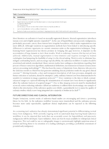
Table 1. Current limitations of radiomics
No. Limitations
1 Limited validation of radiomics models
2 Segmentation required with most literature based on manual segmentation techniques
(1) Operator dependent
(2) Time intensive
(3) Introduces inherent variation that radiomic features may be sensitive to
3 Lack of standardization in the workflow of radiomics
4 Radiomics features are sensitive to variations in imaging protocols, which limits multicentric generalization
Most literature on radiomics is based on manually segmented datasets. Manual segmentation introduces
[22]
human error and is highly operator-dependent . In the case of hepatobiliary and pancreatic malignancies,
particularly pancreatic cancer, these tumors have irregular boundaries, which makes segmentation even
more difficult. Although variations in segmentation methods have been linked to introducing specific
differences in radiomic signatures, no current consensus exists on the segmentation techniques. Deep-
learning-based segmentation has shown promise in bridging this gap; however, it depends on the
accumulation of large datasets to train these models. Work is underway; however, this has not yet been
achieved. Additionally, manual segmentation is time-intensive and requires oversight by trained
radiologists, thus hampering the feasibility of using it in the clinical setting. Third, in order to eliminate bias,
mitigate confounding factors, and encourage reproducibility, the radiomics workflow of studies should be
scrutinized and entirely standardized. Many current studies have ambiguous descriptions regarding their
process of feature extraction algorithms, mathematical definitions, inconsistencies in feature nomenclature
[130]
and pre-processing methodology . This has been the source of skepticism from clinicians who perceive
the radiomics model as a black box that generates satisfying clinical prediction results for any given clinical
[130]
outcome . Moving forwards, a clear and transparent description of all these processes, alongside any
feature reduction or exclusion, should be attempted. Lastly, radiomic features have been demonstrated to be
sensitive to variations in image acquisition protocols [22,131] . Even variations in the time at which contrast-
enhanced images are captured following the administration of contrast have been shown to significantly
affect the radiomic features of acquired images . Due to this, imaging protocols must be standardized
[132]
across all centers evaluating radiomics. Efforts are underway to address these shortcomings, one example of
which is the introduction of the radiomics quality score (RQS), a generalizable tool to assess the quality of
[133]
radiomic studies, which is now being integrated into a majority of studies in the field .
FUTURE DIRECTIONS AND CLINICAL INTEGRATION
Despite limitations, current results of radiomics-based studies are encouraging and denote a promising
future for the field. As the radiomics workflow becomes more standardized and the radiomics process
becomes more easily reproducible, significant clinical implications can be expected in the following
domains.
As a screening tool, radiomics has already demonstrated significant potential in differentiating normal
tissue from malignancy. With currently ongoing work on automated deep learning-based radiomics
workflow, it can be expected that tools that can accurately screen for hepatobiliary and pancreatic
malignancies will be developed. Upon validation, these can then be made available for widespread clinical
integration. This tool could be run on radiographic images to autonomously highlight suspicious areas of
interest and bring them to the attention of a reading radiologist, providing a probability assessment of
potential early malignancy.