
Page 14 - Read Online
P. 14
Scherman. Rare Dis Orphan Drugs J 2023;2:12 https://dx.doi.org/10.20517/rdodj.2023.01 Page 5 of 35
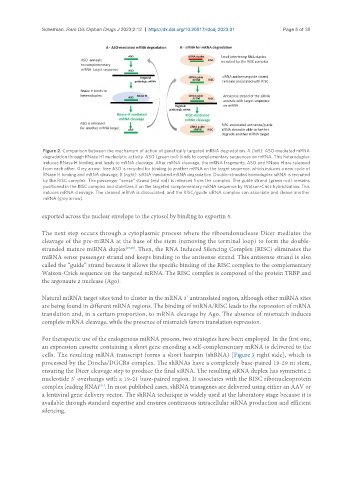
Figure 2. Comparison between the mechanism of action of genetically targeted mRNA degradation. A (left): ASO-mediated mRNA
degradation through RNase H1 nucleolytic activity. ASO (green rod) binds to complementary sequences on mRNA. This heteroduplex
induces RNase H binding and leads to mRNA cleavage. After mRNA cleavage, the mRNA fragments, ASO and RNase H are released
from each other. Grey arrow: free ASO is recycled for binding to another mRNA on the target sequence, which induces a new cycle of
RNase H binding and mRNA cleavage; B (right): siRNA-mediated mRNA degradation. Double-stranded homoduplex siRNA is recruited
by the RISC complex. The passenger “sense” strand (red rod) is released from the complex. The guide strand (green rod) remains
positioned in the RISC complex and stabilizes it on the targeted complementary mRNA sequence by Watson-Crick hybridization. This
induces mRNA cleavage. The cleaved mRNA is dissociated, and the RISC/guide siRNA complex can associate and cleave another
mRNA (grey arrow).
exported across the nuclear envelope to the cytosol by binding to exportin 5.
The next step occurs through a cytoplasmic process where the riboendonuclease Dicer mediates the
cleavage of the pre-miRNA at the base of the stem (removing the terminal loop) to form the double-
stranded mature miRNA duplex [19,20] . Then, the RNA Induced Silencing Complex (RISC) eliminates the
miRNA sense passenger strand and keeps binding to the antisense strand. This antisense strand is also
called the “guide” strand because it allows the specific binding of the RISC complex to the complementary
Watson-Crick sequence on the targeted mRNA. The RISC complex is composed of the protein TRBP and
the argonaute 2 nuclease (Ago).
Natural miRNA target sites tend to cluster in the mRNA 3’ untranslated region, although other miRNA sites
are being found in different mRNA regions. The binding of miRNA/RISC leads to the repression of mRNA
translation and, in a certain proportion, to mRNA cleavage by Ago. The absence of mismatch induces
complete mRNA cleavage, while the presence of mismatch favors translation repression.
For therapeutic use of the endogenous miRNA process, two strategies have been employed. In the first one,
an expression cassette containing a short gene encoding a self-complementary mRNA is delivered to the
cells. The resulting mRNA transcript forms a short hairpin (shRNA) [Figure 3 right side], which is
processed by the Drocha/DGCR8 complex. The shRNAs have a completely base-paired 19-29 nt stem,
ensuring the Dicer cleavage step to produce the final siRNA. The resulting siRNA duplex has symmetric 2
nucleotide 3’ overhangs with a 19-21 base-paired region. It associates with the RISC ribonucleoprotein
complex leading RNAi . In most published cases, shRNA transgenes are delivered using either an AAV or
[21]
a lentiviral gene delivery vector. The shRNA technique is widely used at the laboratory stage because it is
available through standard expertise and ensures continuous intracellular siRNA production and efficient
silencing.