
Page 73 - Read Online
P. 73
Klugmann et al. Rare Dis Orphan Drugs J 2023;2:8 https://dx.doi.org/10.20517/rdodj.2023.05 Page 5 of 9
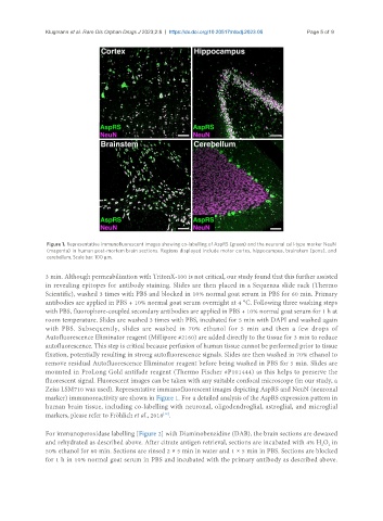
Figure 1. Representative immunofluorescent images showing co-labelling of AspRS (green) and the neuronal cell-type marker NeuN
(magenta) in human post-mortem brain sections. Regions displayed include motor cortex, hippocampus, brainstem (pons), and
cerebellum. Scale bar: 100 µm.
3 min. Although permeabilization with TritonX-100 is not critical, our study found that this further assisted
in revealing epitopes for antibody staining. Slides are then placed in a Sequenza slide rack (Thermo
Scientific), washed 3 times with PBS and blocked in 10% normal goat serum in PBS for 60 min. Primary
antibodies are applied in PBS + 10% normal goat serum overnight at 4 °C. Following three washing steps
with PBS, fluorophore-coupled secondary antibodies are applied in PBS + 10% normal goat serum for 1 h at
room temperature. Slides are washed 3 times with PBS, incubated for 5 min with DAPI and washed again
with PBS. Subsequently, slides are washed in 70% ethanol for 5 min and then a few drops of
Autofluorescence Eliminator reagent (Millipore #2160) are added directly to the tissue for 3 min to reduce
autofluorescence. This step is critical because perfusion of human tissue cannot be performed prior to tissue
fixation, potentially resulting in strong autofluorescence signals. Slides are then washed in 70% ethanol to
remove residual Autofluorescence Eliminator reagent before being washed in PBS for 5 min. Slides are
mounted in ProLong Gold antifade reagent (Thermo Fischer #P101444) as this helps to preserve the
fluorescent signal. Fluorescent images can be taken with any suitable confocal microscope (in our study, a
Zeiss LSM710 was used). Representative immunofluorescent images depicting AspRS and NeuN (neuronal
marker) immunoreactivity are shown in Figure 1. For a detailed analysis of the AspRS expression pattern in
human brain tissue, including co-labelling with neuronal, oligodendroglial, astroglial, and microglial
markers, please refer to Fröhlich et al., 2018 .
[12]
For immunoperoxidase labelling [Figure 2] with Diaminobenzidine (DAB), the brain sections are dewaxed
and rehydrated as described above. After citrate antigen retrieval, sections are incubated with 4% H O in
2
2
50% ethanol for 60 min. Sections are rinsed 2 × 5 min in water and 1 × 5 min in PBS. Sections are blocked
for 1 h in 10% normal goat serum in PBS and incubated with the primary antibody as described above.