
Page 50 - Read Online
P. 50
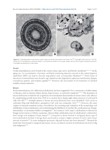
Mu et al. Microstructures 2023;3:2023030  https://dx.doi.org/10.20517/microstructures.2023.05  Page 11 of 21


                Figure 5. Crystal deposition in the urinary system (Reproduced with permission from Evan et al. [150] . Copyright 2005, Elsevier). (A) The
                initial sites of the deposition of kidney stones in transmission electron microscopic images and (B) immunogold staining showed the
                localization of osteopontin in the plaque.


               Ocular
               Ocular mineralization can be found in the cornea, retina, optic nerve, and Bruch’s membrane [92,174-176] . In the
               aging eye, the accumulation of protein- and lipid-containing deposits external to the retinal pigment
               epithelium (RPE) can lead to macular degradation and, consequently, blindness [92,177] . Three types of
               structures of minerals have been found in age-related macular degradation, spherules (whitlockite), plaques
               (amorphous apatite), and nodules (apatite) [20,21] . However, the mechanism of the formation of calcified
               nodules remains unknown.


               Breast
               Breast mineralization, also called microcalcification, has been suggested to be a consequence of either injury
               or diseases, such as chronic kidney disease, hypertension, or metabolic syndrome [178,179] . The formation of
               microcalcification is related to the acquisition of mesenchymal characteristics in breast cancer cells, affected
               by transforming growth factor beta (TGF-β) or nuclear factor kappa-light-chain-enhancer of activated B
               cells (NF-κB) [180,181] . Multiple phases of breast microcalcifications have been identified: CaPs (such as
               carbonate HAp and whitlockite), amorphous CaP, and, less commonly, CaO x [182-185] . However, the exact
               origins of minerals remained unclear. Nevertheless, the screening and evaluation of the morphology and
               distribution of microcalcification aid in determining the likelihood of whether the calcifications are benign,
               intermediate, or necessitate further investigation . For instance, CaOx (type I calcification) is detected in
                                                         [17]
               benign breast lesions or lobular carcinoma, whereas HAp calcification (type II calcification) is detected in
               both benign and malignant breast tissues [18,19] . Compared to those formed in malignant ducts, type II
               microcalcification formed in benign ducts was found to contain a higher amount of CaCO  and a lower
                                                                                              3
                               [186]
                                                                                            [187]
               amount of protein . In some malignant specimens, Mg and Na have also been detected . Elevated Na
               levels have been found in malignant specimens, but no correlation has been found between the level of Mg
               and malignancy .
                             [187]