Page 276 - Read Online
P. 276

Wang et al. Microstructures 2023;3:2023042  https://dx.doi.org/10.20517/microstructures.2023.46  Page 9 of 16


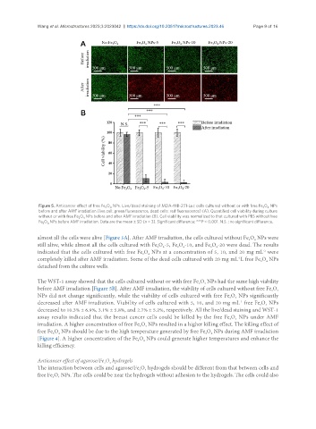
                Figure 5. Anticancer effect of free Fe O  NPs. Live/dead staining of MDA-MB-231-Luc cells cultured without or with free Fe O  NPs
                                         3
                                          4
                                                                                                      4
                                                                                                    3
                before and after AMF irradiation (live cell: green fluorescence, dead cells: red fluorescence) (A). Quantified cell viability during culture
                without or with free Fe O  NPs before and after AMF irradiation (B). Cell viability was normalized to that cultured with PBS without free
                                4
                               3
                Fe O  NPs before AMF irradiation. Data are the mean ± SD (n = 3). Significant difference: ***P < 0.001. N.S. : no significant difference.
                   4
                 3
               almost all the cells were alive [Figure 5A]. After AMF irradiation, the cells cultured without Fe O  NPs were
                                                                                               3
                                                                                                 4
               still alive, while almost all the cells cultured with Fe O -5, Fe O -10, and Fe O -20 were dead. The results
                                                                    3
                                                                                3
                                                              4
                                                            3
                                                                                  4
                                                                      4
                                                                                                    -1
               indicated that the cells cultured with free Fe O  NPs at a concentration of 5, 10, and 20 mg mL  were
                                                         4
                                                       3
               completely killed after AMF irradiation. Some of the dead cells cultured with 20 mg mL L free Fe O  NPs
                                                                                           -1
                                                                                                   3
                                                                                                     4
               detached from the culture wells.
               The WST-1 assay showed that the cells cultured without or with free Fe O  NPs had the same high viability
                                                                            3
                                                                              4
               before AMF irradiation [Figure 5B]. After AMF irradiation, the viability of cells cultured without free Fe O
                                                                                                       3
                                                                                                         4
               NPs did not change significantly, while the viability of cells cultured with free Fe O  NPs significantly
                                                                                       3
                                                                                          4
               decreased after AMF irradiation. Viability of cells cultured with 5, 10, and 20 mg mL  free Fe O  NPs
                                                                                           -1
                                                                                                   3
                                                                                                     4
               decreased to 10.3% ± 6.9%, 3.1% ± 5.8%, and 2.7% ± 5.2%, respectively. All the live/dead staining and WST-1
               assay results indicated that the breast cancer cells could be killed by the free Fe O  NPs under AMF
                                                                                          4
                                                                                       3
               irradiation. A higher concentration of free Fe O  NPs resulted in a higher killing effect. The killing effect of
                                                        4
                                                      3
               free Fe O  NPs should be due to the high temperature generated by free Fe O  NPs during AMF irradiation
                                                                              3
                     3
                                                                                4
                       4
               [Figure 4]. A higher concentration of the Fe O  NPs could generate higher temperatures and enhance the
                                                       4
                                                     3
               killing efficiency.
               Anticancer effect of agarose/Fe O  hydrogels
                                        3
                                          4
               The interaction between cells and agarose/Fe O  hydrogels should be different from that between cells and
                                                     3
                                                        4
               free Fe O  NPs. The cells could be near the hydrogels without adhesion to the hydrogels. The cells could also
                       4
                     3
   271   272   273   274   275   276   277   278   279   280   281