
Page 231 - Read Online
P. 231
Reiss et al. Vessel Plus 2020;4:19 I http://dx.doi.org/10.20517/2574-1209.2020.04 Page 3 of 10
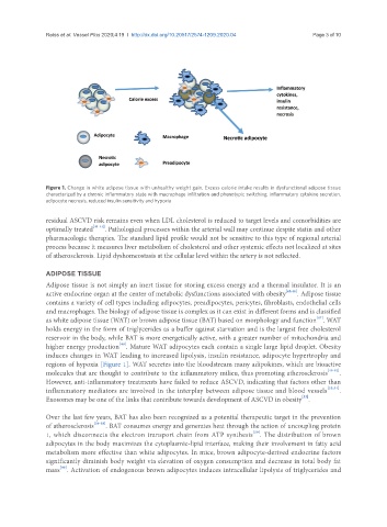
Figure 1. Change in white adipose tissue with unhealthy weight gain. Excess calorie intake results in dysfunctional adipose tissue
characterized by a chronic inflammatory state with macrophage infiltration and phenotypic switching, inflammatory cytokine secretion,
adipocyte necrosis, reduced insulin sensitivity and hypoxia
residual ASCVD risk remains even when LDL cholesterol is reduced to target levels and comorbidities are
optimally treated [41-44] . Pathological processes within the arterial wall may continue despite statin and other
pharmacologic therapies. The standard lipid profile would not be sensitive to this type of regional arterial
process because it measures liver metabolism of cholesterol and other systemic effects not localized at sites
of atherosclerosis. Lipid dyshomeostasis at the cellular level within the artery is not reflected.
ADIPOSE TISSUE
Adipose tissue is not simply an inert tissue for storing excess energy and a thermal insulator. It is an
active endocrine organ at the center of metabolic dysfunctions associated with obesity [45,46] . Adipose tissue
contains a variety of cell types including adipocytes, preadipocytes, pericytes, fibroblasts, endothelial cells
and macrophages. The biology of adipose tissue is complex as it can exist in different forms and is classified
[47]
as white adipose tissue (WAT) or brown adipose tissue (BAT) based on morphology and function . WAT
holds energy in the form of triglycerides as a buffer against starvation and is the largest free cholesterol
reservoir in the body, while BAT is more energetically active, with a greater number of mitochondria and
[48]
higher energy production . Mature WAT adipocytes each contain a single large lipid droplet. Obesity
induces changes in WAT leading to increased lipolysis, insulin resistance, adipocyte hypertrophy and
regions of hypoxia [Figure 1]. WAT secretes into the bloodstream many adipokines, which are bioactive
molecules that are thought to contribute to the inflammatory milieu, thus promoting atherosclerosis [49-52] .
However, anti-inflammatory treatments have failed to reduce ASCVD, indicating that factors other than
inflammatory mediators are involved in the interplay between adipose tissue and blood vessels [53,54] .
[55]
Exosomes may be one of the links that contribute towards development of ASCVD in obesity .
Over the last few years, BAT has also been recognized as a potential therapeutic target in the prevention
of atherosclerosis [56-58] . BAT consumes energy and generates heat through the action of uncoupling protein
[59]
1, which disconnects the electron transport chain from ATP synthesis . The distribution of brown
adipocytes in the body maximizes the cytoplasmic-lipid interface, making their involvement in fatty acid
metabolism more effective than white adipocytes. In mice, brown adipocyte-derived endocrine factors
significantly diminish body weight via elevation of oxygen consumption and decrease in total body fat
[60]
mass . Activation of endogenous brown adipocytes induces intracellular lipolysis of triglycerides and