
Page 187 - Read Online
P. 187
Berezin et al. Vessel Plus 2020;4:15 I http://dx.doi.org/10.20517/2574-1209.2020.03 Page 5 of 13
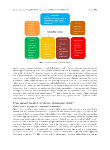
Figure 1. Pleiotropic effects of extracellular vesicles
can be regulated to ensure activation of endothelial cells as well as the attraction and internalization of
various types of circulating blood cells (platelets, mononuclear cells, macrophages, lymphocytes) by the
[42]
endothelial cell surface . Moreover, vascular growth, restoration of vascular integrity and function, as
well as the recruitment of inflammatory cells, may all be directly related to up-regulated expression of
neuregulin-1 in endothelial cells as a result of EV-dependent stimulation, because circulating EVs can be
[43]
a source of a variety of pro-angiogenic mRNAs including neuregulin-1 mRNA . Additionally, EVs may
induce a cytoskeleton-junction response from endothelial cells that is characterized by myosin light chain
phosphorylation, contractile fiber reorganization, VE-cadherin phosphorylation and adherent junction
dissociation. This process is a key mechanism of increasing permeability of the vascular wall, releasing
neutrophil extracellular traps containing citrullinated histones and myeloperoxidase, and in developing
senescence and acceleration of atherosclerosis [44-46] . The proteome of EVs consists of pro-coagulant
components such as tissue factor and phospholipids, which play a pivotal role in coagulation and the
triggering of vasoocclusion in CAD [47,48] .
EVS IN VARIOUS STAGES OF ATHEROSCLEROSIS DEVELOPMENT
Modification of macrophages’ phenotype and function
Macrophages are the primary antigen-presenting cells in atherosclerotic lesions and provide the
fundamental link between microvascular inflammation and atherosclerotic plaque development and
progression. It has been suggested that endothelial cell-derived EVs export microRNA-92a from mother
cells to macrophages in response to atheroprone stimuli to change macrophage phenotype, regulate their
functions and enhance atherosclerotic plaque shaping . Indeed, over-expression of microRNA-92a in
[49]
endothelial cells in atherosclerosis enhances the pro-inflammatory response in the vasculature, supports
low-density lipoprotein (LDL) uptake, and impairs the migration of macrophages through changes in
[50]
their phenotype from atheroprotected to atheroprone . Interestingly, the expression of microRNA-92a
in mature endothelial cells is up-regulated by the combination of several factors such as low shear stress,
atherogenic oxidized LDL, IL-6, atheroprotective Kruppel-like factor (KLF)-2 and KLF-4, and suppressor
of cytokine signaling 5 [51,52] . The expression of pro-inflammatory cytokine-induced markers such as