
Page 188 - Read Online
P. 188
Page 6 of 13 Berezin et al. Vessel Plus 2020;4:15 I http://dx.doi.org/10.20517/2574-1209.2020.03
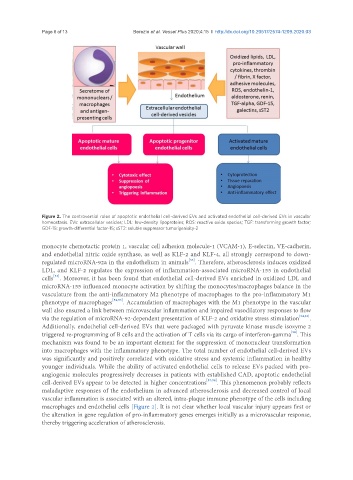
Figure 2. The controversial roles of apoptotic endothelial cell-derived EVs and activated endothelial cell-derived EVs in vascular
homeostasis. EVs: extracellular vesicles; LDL: low-density lipoproteins; ROS: reactive oxide species; TGF: transforming growth factor;
GDF-15: growth-differential factor-15; sST2: soluble suppressor tumorigenisity-2
monocyte chemotactic protein 1, vascular cell adhesion molecule-1 (VCAM-1), E-selectin, VE-cadherin,
and endothelial nitric oxide synthase, as well as KLF-2 and KLF-4, all strongly correspond to down-
[53]
regulated microRNA-92a in the endothelium in animals . Therefore, atherosclerosis induces oxidized
LDL, and KLF-2 regulates the expression of inflammation-associated microRNA-155 in endothelial
[53]
cells . Moreover, it has been found that endothelial cell-derived EVs enriched in oxidized LDL and
microRNA-155 influenced monocyte activation by shifting the monocytes/macrophages balance in the
vasculature from the anti-inflammatory M2 phenotype of macrophages to the pro-inflammatory M1
phenotype of macrophages [52,53] . Accumulation of macrophages with the M1 phenotype in the vascular
wall also ensured a link between microvascular inflammation and impaired vasodilatory responses to flow
via the regulation of microRNA-92-dependent presentation of KLF-2 and oxidative stress stimulation [54,55] .
Additionally, endothelial cell-derived EVs that were packaged with pyruvate kinase muscle isozyme 2
[56]
triggered re-programming of B cells and the activation of T cells via its cargo of interferon-gamma . This
mechanism was found to be an important element for the suppression of mononuclear transformation
into macrophages with the inflammatory phenotype. The total number of endothelial cell-derived EVs
was significantly and positively correlated with oxidative stress and systemic inflammation in healthy
younger individuals. While the ability of activated endothelial cells to release EVs packed with pro-
angiogenic molecules progressively decreases in patients with established CAD, apoptotic endothelial
cell-derived EVs appear to be detected in higher concentrations [57,58] . This phenomenon probably reflects
maladaptive responses of the endothelium in advanced atherosclerosis and decreased control of local
vascular inflammation is associated with an altered, intra-plaque immune phenotype of the cells including
macrophages and endothelial cells [Figure 2]. It is not clear whether local vascular injury appears first or
the alteration in gene regulation of pro-inflammatory genes emerges initially as a microvascular response,
thereby triggering acceleration of atherosclerosis.