
Page 98 - Read Online
P. 98
Orekhov et al. Vessel Plus 2019;3:10 I http://dx.doi.org/10.20517/2574-1209.2019.04 Page 3 of 20
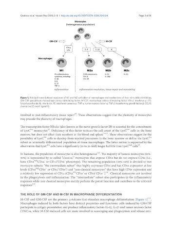
Figure 1. Principal transcriptional regulators of M1 and M2 activation of macrophages and mechanisms of their stimulation/inhibition.
GM-CSF: granulocyte-macrophage colony-stimulating factor; M-CSF: macrophage colony-stimulating factor; IFN-γ: interferon γ; LPS:
lipopolysaccharide; IL: interleukin; IC: interferon consensus; TNF-α: tumor necrosis factor α; TGF-β: transforming growth factor β; CCL18:
chemokine CC-motif ligand 18
[9]
involved in post-inflammatory tissue repair . These observations suggest that the plasticity of monocytes
may precede the plasticity of macrophages.
The transcription factor NR4A1 (also known as the nerve growth factor IB) is essential for the commitment
low
[8]
low
of Ly6C monocytes . Deficiency of this factor reduces the cell count of the Ly6C cells in the bone
marrow, but does not affect their numbers in the blood and spleen [10,11] . These observations suggest for the
low
low
possibility of Ly6C cells to develop from myeloid precursors in the bone marrow or define the Ly6C
subset as terminally differentiated population of tissue macrophages. The latter notion is supported by the
high
[6]
low
observation that Ly6C cells have a significantly (10 to 20-fold) longer half-life than Ly6C cells .
[12]
In humans, the population of monocytes is also heterogeneous . The majority of human monocytes (85%-
90%) is represented by so called “classical” monocytes that express CD14 but do not express CD16 (i.e.,
+
high
-
-
have CD14 CD16 or CD14 CD16 phenotype). The remaining population (10%-15%) is divided to two
monocyte subsets: “the intermediate subset” that highly expresses CD14 and has CD16 expression at low
+
+
+
high
levels (CD14 CD16 or CD14 CD16 ) and “non-classical monocytes” that have high CD16 expression and
+
dim
++ [13]
+
a relatively low expression of CD14 (CD14 CD16 or CD14 CD16 ) . Classical monocytes are involved
in the phagocytosis and inflammation. The “intermediate” subset also participates in the inflammatory
responses while non-classical monocytes mainly perform the patrol function and contribute to the antiviral
[14]
responses .
THE ROLE OF GM-CSF AND M-CSF IN MACROPHAGE DIFFERENTIATION
[15]
M-CSF and GM-CSF are the primary cytokines that stimulate macrophage differentiation [Figure 1] .
Macrophages induced by both factors have distinct properties and functions: cells induced by GM-CSF
participate in antigen presentation and produce inflammatory factors IL-12, IL-23 and tumor necrosis factor
(TNF)-α, while M-CSF-induced cells are more involved in scavenging and phagocytosis and release anti-