
Page 271 - Read Online
P. 271
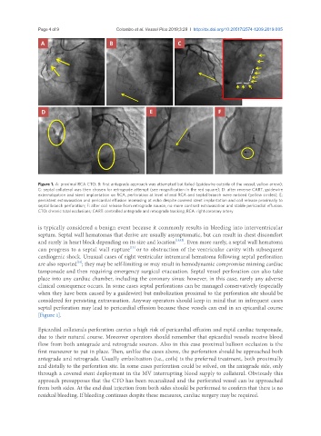
Page 4 of 9                                                 Colombo et al. Vessel Plus 2019;3:29  I  http://dx.doi.org/10.20517/2574-1209.2019.005


               Figure 1. A: proximal RCA CTO; B: first antegrade approach was attempted but failed (guidewire outside of the vessel, yellow arrow);
               C: septal collateral was then chosen for retrograde attempt (see magnification in the red square); D: after reverse CART, guidewire
               externalization and stent implantation on RCA, perforation at level of mid RCA and septal branch were noticed (yellow circles); E:
               persistent extravasation and pericardial effusion increasing at echo despite covered stent implantation and coil release proximally to
               septal branch perforation; F: after coil release from retrograde source, no more contrast extravasation and stable pericardial effusion.
               CTO: chronic total occlusions; CART: controlled antegrade and retrograde tracking; RCA: right coronary artery


               is typically considered a benign event because it commonly results in bleeding into interventricular
               septum. Septal wall hematomas that derive are usually asymptomatic, but can result in chest discomfort
               and rarely in heart block depending on its size and location [11,12] . Even more rarely, a septal wall hematoma
                                                 [13]
               can progress to a septal wall rupture or to obstruction of the ventricular cavity with subsequent
               cardiogenic shock. Unusual cases of right ventricular intramural hematoma following septal perforation
                              [14]
               are also reported ; they may be self-limiting or may result in hemodynamic compromise miming cardiac
               tamponade and then requiring emergency surgical evacuation. Septal vessel perforation can also take
               place into any cardiac chamber, including the coronary sinus: however, in this case, rarely any adverse
               clinical consequence occurs. In some cases septal perforations can be managed conservatively (especially
               when they have been caused by a guidewire) but embolization proximal to the perforation site should be
               considered for persisting extravasation. Anyway operators should keep in mind that in infrequent cases
               septal perforation may lead to pericardial effusion because these vessels can end in an epicardial course
               [Figure 1].

               Epicardial collaterals perforation carries a high risk of pericardial effusion and rapid cardiac tamponade,
               due to their natural course. Moreover operators should remember that epicardial vessels receive blood
               flow from both antegrade and retrograde sources. Also in this case proximal balloon occlusion is the
               first maneuver to put in place. Then, unlike the cases above, the perforation should be approached both
               antegrade and retrograde. Usually embolization (i.e., coils) is the preferred treatment, both proximally
               and distally to the perforation site. In some cases perforation could be solved, on the antegrade side, only
               through a covered stent deployment in the MV interrupting blood supply to collateral. Obviously this
               approach presupposes that the CTO has been recanalized and the perforated vessel can be approached
               from both sides. At the end dual injection from both sides should be performed to confirm that there is no
               residual bleeding. If bleeding continues despite these measures, cardiac surgery may be required.