Page 269 - Read Online
P. 269
Page 2 of 9                                                 Colombo et al. Vessel Plus 2019;3:29  I  http://dx.doi.org/10.20517/2574-1209.2019.005

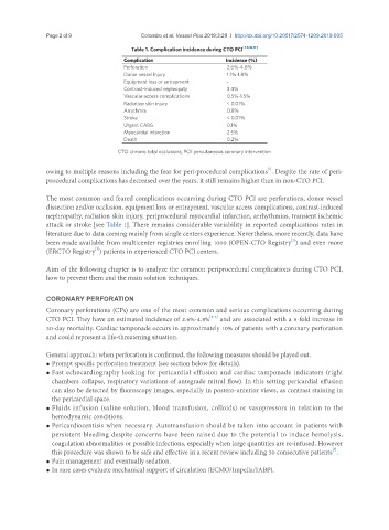
                                         Table 1. Complication incidence during CTO PCI [1-5,16,17]
                                       Complication                   Incidence (%)
                                       Perforation                    2.6%-4.8%
                                       Donor vessel lnjury            1.1%-1.8%
                                       Equipment loss or entrapment   -
                                       Contrast-induced nephropaty    3.4%
                                       Vascular access complications  0.5%-1.5%
                                       Radiation skin injury          < 0.01%
                                       Arrythmia                      0.8%
                                       Stroke                         < 0.01%
                                       Urgent CABG                    0.1%
                                       Myocardial infarction          2.5%
                                       Death                          0.2%
                                     CTO: chronic total occlusions; PCI: percutaneous coronary intervention


                                                                                   [1]
               owing to multiple reasons including the fear for peri-procedural complications . Despite the rate of peri-
               procedural complications has decreased over the years, it still remains higher than in non-CTO PCI.

               The most common and feared complications occurring during CTO PCI are perforations, donor vessel
               dissection and/or occlusion, equipment loss or entrapment, vascular access complications, contrast-induced
               nephropathy, radiation skin injury, periprocedural myocardial infarction, arrhythmias, transient ischemic
               attack or stroke [see Table 1]. There remains considerable variability in reported complications rates in
               literature due to data coming mainly from single centers experience. Nevertheless, more recently, data have
                                                                                          [2]
               been made available from multicenter registries enrolling 1000 (OPEN-CTO Registry ) and even more
                              [3]
               (ERCTO Registry ) patients in experienced CTO PCI centers.
               Aim of the following chapter is to analyze the common periprocedural complications during CTO PCI,
               how to prevent them and the main solution techniques.


               CORONARY PERFORATION
               Coronary perforations (CPs) are one of the most common and serious complications occurring during
                                                                 [2-6]
               CTO PCI. They have an estimated incidence of 2.6%-4.8%  and are associated with a 5-fold increase in
               30-day mortality. Cardiac tamponade occurs in approximately 10% of patients with a coronary perforation
               and could represent a life-threatening situation.

               General approach: when perforation is confirmed, the following measures should be played out.
               • Prompt specific perforation treatment (see section below for details).
               • Fast echocardiography looking for pericardial effusion and cardiac tamponade indicators (right
                 chambers collapse, respiratory variations of antegrade mitral flow). In this setting pericardial effusion
                 can also be detected by fluoroscopy images, especially in postero-anterior views, as contrast staining in
                 the pericardial space.
               • Fluids infusion (saline solution, blood transfusion, colloids) or vasopressors in relation to the
                 hemodynamic conditions.
               • Pericardiocentisis when necessary. Autotransfusion should be taken into account in patients with
                 persistent bleeding despite concerns have been raised due to the potential to induce hemolysis,
                 coagulation abnormalities or possible infections, especially when large quantities are re-infused. However
                                                                                                       [7]
                 this procedure was shown to be safe and effective in a recent review including 30 consecutive patients .
               • Pain management and eventually sedation.
               • In rare cases evaluate mechanical support of circulation (ECMO/Impella/IABP).

