Page 230 - Read Online
P. 230
Page 6 of 9                                          Van der Merwe et al. Vessel Plus 2019;3:24  I  http://dx.doi.org/10.20517/2574-1209.2019.17


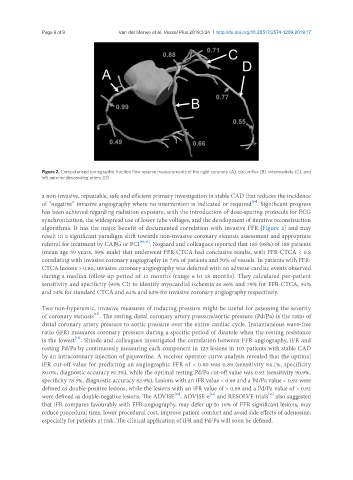
               Figure 2. Computerised tomographic fraction flow reserve measurements of the right coronary (A), circumflex (B), intermediate (C), and
               left anterior descending artery (D)

               a non-invasive, repeatable, safe and efficient primary investigation in stable CAD that reduces the incidence
                                                                                      [39]
               of “negative” invasive angiography where no intervention is indicated or required . Significant progress
               has been achieved regarding radiation exposure, with the introduction of dose-sparing protocols for ECG
               synchronization, the widespread use of lower tube voltages, and the development of iterative reconstruction
               algorithms. It has the major benefit of documented correlation with invasive FFR [Figure 2] and may
               result in a significant paradigm shift towards non-invasive coronary stenosis assessment and appropriate
               referral for treatment by CABG or PCI [40,41] . Nogaard and colleagues reported that 185 (98%) of 189 patients
               (mean age 59 years, 59% male) that underwent FFR-CTCA had conclusive results, with FFR-CTCA < 0.8
               correlating with invasive coronary angiography in 73% of patients and 70% of vessels. In patients with FFR-
               CTCA lesions > 0.80, invasive coronary angiography was deferred with no adverse cardiac events observed
               during a median follow-up period of 12 months (range 6 to 18 months). They calculated per-patient
               sensitivity and specificity (95% CI) to identify myocardial ischemia as 86% and 79% for FFR-CTCA, 94%
               and 34% for standard CTCA and 64% and 83% for invasive coronary angiography respectively.


               Two non-hyperemic, invasive measures of inducing pressure might be useful for assessing the severity
                                 [42]
               of coronary stenosis . The resting distal coronary artery pressure/aortic pressure (Pd/Pa) is the ratio of
               distal coronary artery pressure to aortic pressure over the entire cardiac cycle. Instantaneous wave-free
               ratio (iFR) measures coronary pressure during a specific period of diastole when the resting resistance
                          [42]
               is the lowest . Shiode and colleagues investigated the correlation between FFR-angiography, iFR and
               resting Pd/Pa by continuously measuring each component in 123 lesions in 103 patients with stable CAD
               by an intracoronary injection of papaverine. A receiver operator curve analysis revealed that the optimal
               iFR cut-off value for predicting an angiographic FFR of < 0.80 was 0.89 (sensitivity 84.1%, specificity
               80.0%, diagnostic accuracy 81.3%), while the optimal resting Pd/Pa cut-off value was 0.92 (sensitivity 90.9%,
               specificity 78.5%, diagnostic accuracy 82.9%). Lesions with an iFR value < 0.89 and a Pd/Pa value < 0.92 were
               defined as double-positive lesions, while the lesions with an iFR value of > 0.89 and a Pd/Pa value of > 0.92
                                                            [43]
               were defined as double-negative lesions. The ADVISE , ADVISE-e  and RESOLVE trials  also suggested
                                                                                           [45]
                                                                        [44]
               that iFR compares favourably with FFR-angiography, may defer up to 16% of FFR significant lesions, may
               reduce procedural time, lower procedural cost, improve patient comfort and avoid side effects of adenosine,
               especially for patients at risk. The clinical application of iFR and Pd/Pa will soon be defined.

