
Page 227 - Read Online
P. 227
Van der Merwe et al. Vessel Plus 2019;3:24 I http://dx.doi.org/10.20517/2574-1209.2019.17 Page 3 of 9
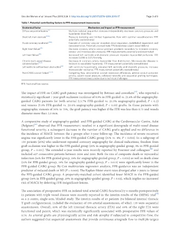
Table 1. Potential contributing factors to FFR measurement inaccuracies
Anatomical factor Mechanism and Impact on FFR-measurement
Diffuse sequential lesions [5] Multiple isolated sequential stenoses independently decrease coronary pressure and
hyperaemic blood flow
Short left main stenosis [15,16] Pressure damping and limited hyperaemic flow with optimal vasodilatation. FFR
potentially overestimated
Acute coronary syndrome [17,18] Cascade of coronary vascular receptors down-regulation, endothelial impairment and
vasoconstriction. Potentially overestimate FFR/deleterious culprit-vessel deferral
Right heart failure [19] Decrease coronary arterio-venous pressure gradients secondary to increases coronary
venous- and microvascular pressures. FFR measurements potentially underestimated
Left heart failure [19] Increased left ventricle end-diastolic pressure impedes myocardial perfusion. FFR
increases 0.008 to 0.01/1 mmHg
Chronic multi-vessel disease Decrease in coronary artery-myocardial flow distribution. Microvascular disease is
collateralization [20,21] resistant to vasodilator hyperemia. FFR measurement potentially overestimated
Left ventricle outflow tract obstruction [22] Left ventricle hypertrophy, elevated left ventricle end-diastolic pressure, increase
microvascular resistance. FFR measurement potentially overestimated
Post-CABG conduit failure [23,24] Competing flow, veno-arterial conduit resistance differences, arterial conduit autocrine
activity, culprit-vessel pressure, collateral networks and sequential grafting techniques.
FFR measurement inaccuracies due to technical challenges
FFR: fractional flow reserve
[33]
The impact of FFR on CABG graft patency was investigated by Botman and coworkers , who reported a
statistically significant 1 year graft occlusion incidence of 8.9% in FFR-guided vs. 21.4% of the angiography-
guided CABG patients for both arterial (13.7% FFR-guided vs. 21.9% angiography-guided; P < 0.2)
and venous (5.9% FFR-guided vs. 20.0% angiography-guided; P < 0.03) grafts. In those patients with
angiographic stenosis of 50% to 70%, the graft patency was higher if the FFR was less than 0.75 and vessels
diameter more than 2.0 mm.
A comparative study of angiography-guided- and FFR-guided CABG at the Cardiovascular Centre, Aalst
[33]
(Belgium) observed that FFR measurement resulted in a significant downgrade of multi-vessel disease
functional severity, a subsequent decrease in the number of CABG grafts applied and no difference in
the incidence of MACE between the 2 groups after 3-year follow-up. The incidence of severe recurrent
angina was significantly lower in the FFR-guided CABG group (31% vs. 4%; P < 0.001). In a subgroup of
155 patients (25%) who underwent repeated coronary angiography for clinical indications, freedom from
graft occlusion was higher in the FFR-guided group (21% in angiography-guided group, 5% in FFR-guided
[34]
group, P = 0.031). The extended 6-year results were recently reported by Fournier and colleagues and
included 627 consecutive patients between 2006 and 2010. Both the rate of composite death or myocardial
infarction (16% for FFR-guided group, 25% for angiography-guided group, P = 0.020) as well as death alone
(11% for FFR-guided group, 18% for angiography-guided group, P = 0.013) were significantly lower in the
FFR-guided CABG group. By Cox multivariate regression analysis, FFR-guidance was an independent
predictor of reduced death or MI (P = 0.008). The Kaplan-Meier event rates diverged after 3 years to favour
the FFR-guided CABG group. A propensity-matched cohort identified fewer MACE in the FFR-guided
group (16% in FFR-guided group, 25% in angiographic-guided group, P < 0.02), which implies no increased
risk of MACE by deferring FFR insignificant lesions.
The association of preoperative FFR on isolated total arterial CABG functionality 6 months postoperatively
in patients with triple vessel disease were recently reported in the interim results of the IMPAG trial [35]
as a 2-centre, single-arm, blinded study. The interim results of 63 patients (54 bilateral internal thoracic
Y-graft configurations), included the evaluation of 199 arterial anastomoses, of which 135 were sequential
anastomoses. Overall, 85% of the left internal thoracic artery (ITA) and 69% of the right ITA were
functional and patent, which was statistically significantly associated with preoperative FFR values of
0.78. As arterial grafts are physiologically active and risk atrophy if subjected to competitive flow, the
authors suggested that sequential anastomosis that provide continuous antegrade flow to multiple targets