Page 392 - Read Online
P. 392
Page 4 of 6                                             Lopez-Marco et al. Vessel Plus 2018;2:40  I  http://dx.doi.org/10.20517/2574-1209.2018.67


                                       A


                                       B


                                       C

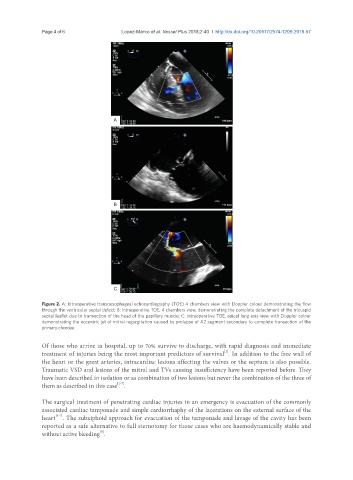
               Figure 2. A: Intraoperative transoesophageal echocardiography (TOE) 4 chambers view with Doppler colour demonstrating the flow
               through the ventricular septal defect; B: intraoperative TOE, 4 chambers view, demonstrating the complete detachment of the tricuspid
               septal leaflet due to transection of the head of the papillary muscle; C: intraoperative TOE, apical long axis view with Doppler colour
               demonstrating the eccentric jet of mitral regurgitation caused by prolapse of A2 segment secondary to complete transection of the
               primary chordae


               Of those who arrive in hospital, up to 70% survive to discharge, with rapid diagnosis and immediate
                                                                             [2]
               treatment of injuries being the most important predictors of survival . In addition to the free wall of
               the heart or the great arteries, intracardiac lesions affecting the valves or the septum is also possible.
               Traumatic VSD and lesions of the mitral and TVs causing insufficiency have been reported before. They
               have been described in isolation or as combination of two lesions but never the combination of the three of
                                         [1-7]
               them as described in this case .

               The surgical treatment of penetrating cardiac injuries in an emergency is evacuation of the commonly
               associated cardiac tamponade and simple cardiorrhaphy of the lacerations on the external surface of the
                    [1-7]
               heart . The subxiphoid approach for evacuation of the tamponade and lavage of the cavity has been
               reported as a safe alternative to full sternotomy for those cases who are haemodynamically stable and
                                   [8]
               without active bleeding .

