Page 393 - Read Online
P. 393
Lopez-Marco et al. Vessel Plus 2018;2:40  I  http://dx.doi.org/10.20517/2574-1209.2018.67                                           Page 5 of 6


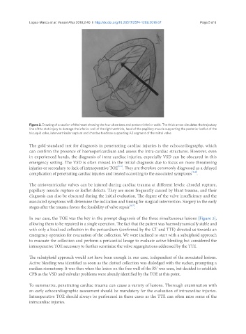
               Figure 3. Drawing of a section of the heart showing the four chambers and postero-inferior walls. The thick arrow simulates the trajectory
               line of the stab injury to damage the inferior wall of the right ventricle, head of the papillary muscle supporting the posterior leaflet of the
               tricuspid valve, interventricular septum and chordae tendinae supporting A2 segment of the mitral valve

               The gold-standard test for diagnosis in penetrating cardiac injuries is the echocardiography, which
               can confirm the presence of haemopericardium and assess the intra-cardiac structures. However, even
               in experienced hands, the diagnosis of intra-cardiac injuries, especially VSD can be obscured in this
               emergency setting. The VSD is often missed in the initial diagnosis due to focus on more threatening
                                                          [1-5]
               injuries or secondary to lack of intraoperative TOE . They are therefore commonly diagnosed as a delayed
                                                                                              [1-6]
               complication of penetrating cardiac injuries and treated according to the associated symptoms .
               The atrioventricular valves can be injured during cardiac trauma at different levels: chordal rupture,
               papillary muscle rupture or leaflet defects. They are more frequently caused by blunt trauma, and their
               diagnosis can also be obscured during the initial evaluation. The degree of the valve insufficiency and the
               associated symptoms will determine the indication and timing for surgical intervention. Surgery in the early
                                                                [4-7]
               stages after the trauma favors the feasibility of valve repair .

               In our case, the TOE was the key in the prompt diagnosis of the three simultaneous lesions [Figure 3],
               allowing them to be repaired in a single operation. The fact that the patient was haemodynamically stable and
               with only a localized collection in the pericardium (confirmed by the CT and TTE) directed us towards an
               emergency operation for evacuation of the collection. We were inclined to start with a subxiphoid approach
               to evacuate the collection and perform a pericardial lavage to evaluate active bleeding but considered the
               intraoperative TOE necessary to further scrutinize the valve regurgitations addressed by the TTE.


               The subxiphoid approach would not have been enough in our case, independent of the associated lesions.
               Active bleeding was identified as soon as the clotted collection was dislodged with the sucker, prompting a
               median sternotomy. It was then when the lesion on the free wall of the RV was seen, but decided to establish
               CPB as the VSD and valvular problems were already identified by the TOE at this point.

               To summarize, penetrating cardiac trauma can cause a variety of lesions. Thorough examination with
               an early echocardiographic assessment should be mandatory for the evaluation of intracardiac injuries.
               Intraoperative TOE should always be performed in these cases as the TTE can often miss some of the
               intracardiac injuries.

