Page 383 - Read Online
P. 383
Page 6 of 11                                                    Elsayed et al. Vessel Plus 2018;2:39  I  http://dx.doi.org/10.20517/2574-1209.2018.65


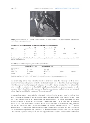
               Figure 1. Representative image of CT coronary angiography showing left internal mammary artery (LIMA) patch with patent LIMA-left
               anterior descending artery anastomosis

               Table 4. Comparison between pre and postoperative New York Heart Association class

                                  Preoperative (n = 30)    Postoperative (n = 29)
               NYHA class                                                             Z            P
                                 No.           %           No.           %
               I                 0            0.0          20           69.0
               II                15           50.0         9            31.0        4.919*       < 0.001*
               III               15           50.0         0            0.0
               *Statistically significant at P ≤ 0.05; Z and P values for Wilcoxon signed ranks test for comparing between pre and postoperative; NYHA:
               New York Heart Association


               Table 5. Comparison between pre and postoperative ejection fraction
                                   Preoperative (n = 30)  Postoperative (n = 29)  T              P
               Ejection fraction
                 Min-Max             35.0-70.0             48.0-68.0
                 Mean ± SD           51.23 ± 9.02          56.28 ± 5.62         4.968*         < 0.001*
                 Median              49.0                  58.0
               *Statistically significant at P ≤ 0.05; T and P values for Paired t-test for comparing between pre and postoperative

               Endarterectomy entails removal of the atherosclerotic core from the coronary vessel. In closed
               endarterectomy (also called pull out method) a small arteriotomy is used to dissect the atherosclerotic plaque
               out of the coronary vessel by using steady and gentle traction. It’s a simpler technique but was criticized due
               to the possibility of occlusion of the distal LAD or its branches by insufficient endarterectomy the so called
                             [7]
               snowplow effect . In addition blind traction on the atherosclerotic core can lead to tears and iatrogenic
               intimal flaps that would lead to occlusion of the coronary vessel .
                                                                    [8]
               In open endarterectomy a longitudinal arteriotomy is performed on the coronary vessel beyond the limits
               of the atheromatous plaque and the atherosclerotic plaque is dissected under vision from the coronary vessel
               and its side branches avoiding any residual obstruction and repairing any intimal flaps that might occur
               during the removal of the plaque. The coronary is then reconstructed using an onlay patch of saphenous
               vein or LIMA itself. Advocates of coronary reconstruction using the saphenous vein patch suggested
                                                                                               [6,8]
               that it is very easy to use, easy to harvest and enlarged the lumen of the reconstructed LAD . However
               others accused it of being more time consuming, as you convert one anastomosis into two anastomoses,
               first anastomosis of saphenous vein graft (SVG) to the endarterectomized LAD, then anastomosing the
               LIMA onto the saphenous vein patch. It was also suggested that this complex vascular bed formed of three

