
Page 247 - Read Online
P. 247
Depboylu et al. Vessel Plus 2018;2:26 I http://dx.doi.org/10.20517/2574-1209.2018.39 Page 7 of 11
A B
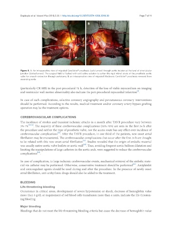
Figure 4. A: An intraoperative view of migrated CoreValve® prosthesis (solid arrow) through aortic incision at the level of sino-tubular
junction (dotted arrow). The surgical field is flushed with cold saline solution to soften the rigid nitinol struts of the prosthetic aortic
valve for smooth extraction through aortotomy; B: an intraoperative view of migrated Medtronic-CoreValve® prosthesis removed from
ascending aorta
(particularly CK-MB) in the post-procedural 72 h, detection of the loss of viable myocardium on imaging
[3]
and ventricular wall motion abnormality also indicate the peri-procedural myocardial infarction .
In case of such complications, selective coronary angiography and percutaneous coronary interventions
should be performed. According to the results, medical treatment and/or coronary artery bypass grafting
operation may be the treatment options.
CEREBROVASCULAR COMPLICATIONS
The incidence of strokes and transient ischemic attacks in a month after TAVR procedure vary between
3%-7% [30,31] . The majority of these cerebrovascular complications (50%-70%) are seen in the first 24 h after
the procedure and neither the type of prosthetic valve, nor the access route has any effect over incidence of
[20]
cerebrovascular complications . After the TAVR procedure, in one third of the patients, new onset atrial
fibrillation may be encountered. The cerebrovascular complications that occur after the first 24 h are thought
[32]
to be related with this new onset atrial fibrillation . Studies revealed that the origin of embolic material
[33]
was usually native aortic valve leaflets or aortic wall . Thus, avoiding frequent aortic balloon dilatation and
limiting the manipulations of large catheters in the aortic arch, were suggested to reduce the cerebrovascular
[34]
complications .
In case of complication, in large ischemic cerebrovascular events, mechanical retrieval of the embolic mate-
[14]
rial via catheter may be performed. Otherwise, conservative treatment should be performed . Antiplatelet
and anticoagulant agents should be used during and after the procedure. In the presence of newly onset
atrial fibrillation, anti-arrhythmic drugs should also be added to the treatment.
BLEEDING
Life-threatening bleeding
Occurrence in critical areas, development of severe hypotension or shock, decrease of hemoglobin value
more than 5 g/dL or requirement of red blood cells transfusion more than 4 units, indicate the life-threaten-
ing bleeding.
Major bleeding
Bleedings that do not meet the life-threatening bleeding criteria but cause the decrease of hemoglobin value