
Page 246 - Read Online
P. 246
Page 6 of 11 Depboylu et al. Vessel Plus 2018;2:26 I http://dx.doi.org/10.20517/2574-1209.2018.39
A B
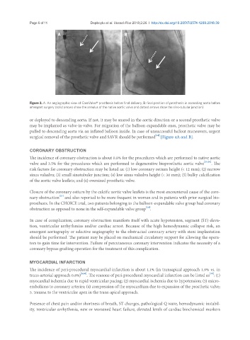
Figure 3. A: An angiographic view of CoreValve® prosthesis before final delivery; B: final position of prosthesis in ascending aorta before
emergent surgery (solid arrows show the annulus of the native aortic valve and dotted arrows show the sino-tubular junction)
or deployed to descending aorta. If not, it may be snared in the aortic direction or a second prosthetic valve
may be implanted as valve-in-valve. For migration of the balloon expandable ones, prosthetic valve may be
pulled to descending aorta via an inflated balloon inside. In case of unsuccessful bailout maneuvers, urgent
[14]
surgical removal of the prosthetic valve and SAVR should be performed [Figure 4A and B].
CORONARY OBSTRUCTION
The incidence of coronary obstruction is about 0.8% for the procedures which are performed to native aortic
valve and 3.5% for the procedures which are performed to degenerative bioprosthetic aortic valve [25,26] . The
risk factors for coronary obstruction may be listed as: (1) low coronary ostium height (< 12 mm); (2) narrow
sinus valsalva; (3) small sinotubuler junction; (4) low sinus valsalva height (< 30 mm); (5) bulky calcification
of the aortic valve leaflets; and (6) oversized prosthetic valve.
Closure of the coronary ostium by the calcific aortic valve leaflets is the most encountered cause of the coro-
[27]
nary obstruction and also reported to be more frequent in women and in patients with prior surgical bio-
prosthesis. In the CHOICE trial, two patients belonging to the balloon-expandable valve group had coronary
[28]
obstruction as opposed to none in the self-expandable valve group .
In case of complication; coronary obstruction manifests itself with acute hypotension, segment (ST) eleva-
tion, ventricular arrhythmias and/or cardiac arrest. Because of the high hemodynamic collapse risk, an
emergent aortography or selective angiography to the obstructed coronary artery with stent implantation
should be performed. The patient may be placed on mechanical circulatory support for allowing the opera-
tors to gain time for intervention. Failure of percutaneous coronary intervention indicates the necessity of a
coronary bypass grafting operation for the treatment of this complication.
MYOCARDIAL INFARCTION
The incidence of peri-procedural myocardial infarction is about 1.1% (in transapical approach 1.9% vs. in
[29]
trans-arterial approach 0.8%) [8,20] . The reasons of peri-procedural myocardial infarction can be listed as : (1)
myocardial ischemia due to rapid ventricular pacing; (2) myocardial ischemia due to hypotension; (3) micro-
embolisms to coronary arteries; (4) compression of the myocardium due to expansion of the prosthetic valve;
5. trauma to the ventricular apex in the trans-apical approach.
Presence of chest pain and/or shortness of breath, ST changes, pathological Q wave, hemodynamic instabil-
ity, ventricular arrhythmia, new or worsened heart failure, elevated levels of cardiac biochemical markers