
Page 59 - Read Online
P. 59
Page 4 of 12 Bui et al. Vessel Plus 2021;6:31 https://dx.doi.org/10.20517/2574-1209.2021.97
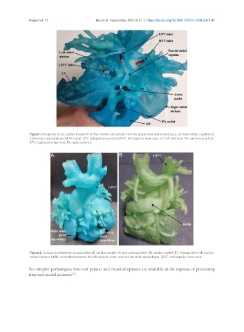
Figure 1. Preoperative 3D cardiac model of the five-month-old patient from the apical view demonstrating a common atrium, ipsilateral
pulmonary veins and partial AV canal. LPV: Left pulmonary vein; LSVC: left superior vena cava; LV: left ventricle; PA: pulmonary artery;
RPV: right pulmonary vein; RV: right ventricle.
Figure 2. Comparison between preoperative 3D cardiac model (A) and postoperative 3D cardiac model (B). Postoperative 3D cardiac
model shows a baffle connection between the left superior vena cava and the atrial appendages. LSVC: Left superior vena cava.
For simpler pathologies, low-cost printer and material options are available at the expense of processing
[25]
time and model accuracy .