
Page 191 - Read Online
P. 191
Page 4 of 11 Misra et al. Vessel Plus 2022;6:14 https://dx.doi.org/10.20517/2574-1209.2021.89
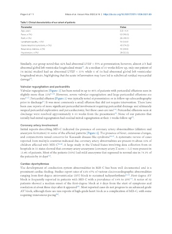
Table 1. Clinical characteristics of our cohort of patients
Parameter Value
Age, years 6.8 ± 4.4
Fever, n (%) 53 (98.0)
Rash, n (%) 26 (48.0)
Lymphadenopathy, n (%) 13 (24.0)
Gastrointestinal symptoms, n (%) 40 (74.0)
Respiratory distress, n (%) 15 (28.0)
Hypotension, n (%) 28 (52.0)
Similarly, our group noted that 42% had abnormal LVEF < 55% at presentation; however, almost 2/3 had
abnormal global left ventricular longitudinal strain . At a median of 10 weeks follow up, only one patient of
[7]
54 initial studied had an abnormal LVEF < 55% while 6 of 54 had abnormal global left ventricular
longitudinal strain, highlighting that the acute inflammation may have led to subclinical residual myocardial
damage .
[7]
Valvular regurgitation and pericarditis
Valvular regurgitation [Figure 1] has been noted in up to 40% of patients with pericardial effusions seen in
slightly more than 25% [13,18] . However, severe valvular regurgitation and large pericardial effusions are
rare [7,13] . Pericardial effusion [Figure 2] was typically noted at presentation or in follow-up echocardiography
prior to discharge . It was most commonly a small effusion that did not require intervention. There have
[7]
been case reports of more significant pericardial involvement requiring pericardial drainage and ultimately
[19]
surgical pericardial exploration and pericardiectomy, but these cases are rare . Pericardial effusions seen at
[7]
discharge were resolved approximately 8-10 weeks from the presentation . None of our patients that
initially had mitral regurgitation had residual mitral regurgitation at their 3 weeks follow-up .
[7]
Coronary artery involvement
Initial reports describing MIS-C indicated the presence of coronary artery abnormalities (dilation and
aneurysm formation) in some of the affected patients [Figure 3]. The presence of fever, cutaneous changes,
and conjunctivitis raised concerns for Kawasaki disease-like syndrome [20,21] . A systematic review of cases
reported from multiple countries indicated that coronary artery abnormalities are present in about 20% of
children affected with MIS-C [21,22] . A large study in the United States involving data collection from 66
hospitals in 31 states showed that coronary artery aneurysms (coronary artery Z score > 2.5) were present in
13.4% of patients. Most of the patients (93%) had mild aneurysms that regressed to normal size in 79.1% of
[12]
the patients by 30 days .
Cardiac dysrhythmias
The development of conduction system abnormalities in MIS-C has been well documented and is a
prominent cardiac finding. Studies report rates of 12%-67% of various electrocardiographic abnormalities
ranging from first-degree atrioventricular (AV) block to sustained tachyarrhythmia [22,23] . First-degree AV
[24]
block is frequently reported in patients with MIS-C with a prevalence of 19% to 25% . A series of 32
patients showed a median onset of the first-degree block at 8 days from the start of symptoms and
resolution at about three days after it appeared . Most reported cases do not progress to an advanced grade
[25]
AV block, although there are rare reports of high-grade heart block as a complication of MIS-C, with some
requiring transvenous pacing .
[26]