
Page 194 - Read Online
P. 194
Misra et al. Vessel Plus 2022;6:14 https://dx.doi.org/10.20517/2574-1209.2021.89 Page 7 of 11
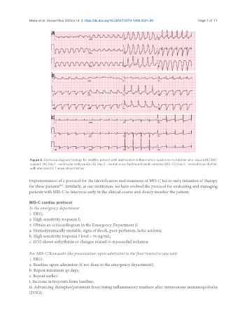
Figure 5. Electrocardiogram findings for another patient with multisystem inflammatory syndrome in children who required ECMO
support. (A) Day 1 - ventricular tachycardia. (B) Day 2 - normal sinus rhythm with wide complex QRS. (C) Day 5 - normal sinus rhythm
with non-specific T wave abnormalities.
implementation of a protocol for the identification and treatment of MIS-C led to early initiation of therapy
[27]
for these patients . Similarly, at our institution, we have evolved the protocol for evaluating and managing
patients with MIS-C to intervene early in the clinical course and closely monitor the patient.
MIS-C cardiac protocol
In the emergency department
1. EKG;
2. High sensitivity troponin I;
3. Obtain an echocardiogram in the Emergency Department if:
a. Hemodynamically unstable, signs of shock, poor perfusion, lactic acidosis;
b. High sensitivity troponin I level > 50 ng/mL;
c. ECG shows arrhythmia or changes related to myocardial ischemia.
For MIS-C/Kawasaki-like presentation: upon admission to the floor/intensive care unit
1. EKG:
a. Baseline: upon admission (if not done in the emergency department);
b. Repeat minimum q3 days;
c. Repeat earlier:
i. Increase in troponin from baseline;
ii. Advancing therapies/persistent fever/rising inflammatory markers after intravenous immunoglobulin
(IVIG);